$410.00
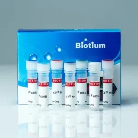
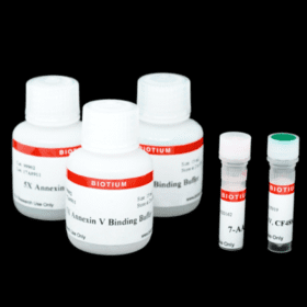
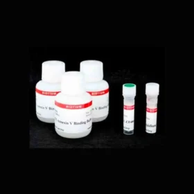
NucView®488 and RedDot™ Apoptosis/Necrosis Assay Kit contains NucView®488 Caspase-3 Substrate for detection of caspase-3/7 activity. The far-red dead-cell stain RedDot™ is included for staining necrotic and late apoptotic cells that have compromised plasma membrane integrity. This kit provides a convenient tool for profiling apoptotic and necrotic cell populations by fluorescence microscopy or flow cytometry.
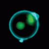
In contrast to other fluorogenic caspase substrates or fluorescent caspase inhibitor based (FLICA) assays, NucView®488 Caspase-3 Substrate can be used to detect caspase-3/7 activity within individual intact cells without inhibiting apoptosis progression. The substrate consists of a fluorogenic DNA dye coupled to the caspase-3/7 DEVD recognition sequence. The substrate, which is initially non-fluorescent, penetrates the plasma membrane and enters the cytoplasm. In apoptotic cells, caspase-3/7 cleaves the substrate, releasing the high-affinity DNA dye, which migrates to the cell nucleus and stains DNA with bright green fluorescence. Thus, NucView®488 Caspase-3 Substrate allows detection caspase-3/7 activity and visualization of morphological changes in the nucleus during apoptosis.
RedDot™ is a cell membrane-impermeable, far-red dye with high selectivity for membrane compromised or dead cells. RedDot™ has far-red emission at 695 nm for detection in the Cy®5 channel, well-separated from the green fluorescence of NucView®488. The excitation maximum of RedDot™ is 665 nm, but it can be efficiently excited by wavelengths from 488 to 647 nm, and therefore can be used with the 488 nm flow cytometry laser line.
Note: while NucView®488 staining is formaldehyde-fixable and compatible with subsequent immunostaining, fixation after staining with RedDot™ is not recommended because it can result in increased background staining of healthy cells.
NucView® enzyme substrate technology is covered by U.S. patents.
Stopa, Kinga B et al.
Driver Mutations of Pancreatic Cancer Affect Ca2+ Signaling and ATP Production
Function (Oxford, England) vol. 4,5 zqad035.
Article Snippet: “Matrigel, growth factor reduced, phenol red-free, was purchased from Corning, USA; NucView 488 & RedDot 2 Apoptosis and Necrosis Kit was purchased from Biotium, USA; TAE buffer was purchased from Bio-Rad, USA; and Thapsiargin was purchased from Cayman, USA.”
Swain, Risa Mia et al.
Thiophene derivative inflicts cytotoxicity via an intrinsic apoptotic pathway on human acute lymphoblastic leukemia cells
PloS one vol. 18,12 e0295441.
DOI: 10.1371/journal.pone.0295441
Article Snippet: “Reference Part NucView® 488 & RedDot™ 2 Apoptosis & Necrosis Kit—Biotium.“
Amalia Sintou, Sarah el Rifai, Catherine Mansfield, et al.
Persistent anti-heart autoimmunity causes cardiomyocyte damage in chronic heart failure
bioRxiv 542597;
DOI: https://doi.org/10.1101/542597
Article Snippet: “Apoptosis assays were performed using Nucleocounter NC-3000 and a Flexicyte apoptosis/necrosis detection kit based on staining with caspase 3 substrate NucView 488 and RedDot 2 (all Biotium, Cambridge Bioscience, UK).”
Anuradha Vajjala, Debabrata Biswas, Kelvin Kian Long Chong, Wei Hong Tay, Emanuel Hanski, Kimberly A Kline.
Streptolysin-induced endoplasmic reticulum stress promotes group A streptococcal in vivo biofilm formation and necrotizing fasciitis
bioRxiv 183012;
DOI: https://doi.org/10.1101/183012
Article Snippet: “ See supplementary methods for additional details. Cell death was also assessed using a NucView 488 and RedDot 2 Apoptosis and Necrosis Kit (Biotium, California, USA).”
Contact our Customer Care, Sales & Scientific Assistance
Consult and asked questions about our products & services
Documentation of Technical & Safety Data Sheet, Guides and more...
We gladly support you by keeping you updated on our latest products and the developments around our services.
362 Upper Paya Lebar Rd, #07-15,
Singapore 534963
© 2015-2023 Atlantis Bioscience Pte Ltd. All rights reserved. Co Reg No: 201539516N