- Your cart is empty
- Continue Shopping


Blue fluorescent caspase-3/7 substrate for detecting apoptosis in intact cells by confocal microscopy, flow cytometry, or live cell imaging.
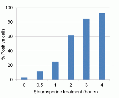
NucView® 405 Caspase-3 Substrate is a convenient tool for detecting apoptosis in intact cells based on caspase-3/7 activity using confocal microscopy, flow cytometry, or live cell imaging.
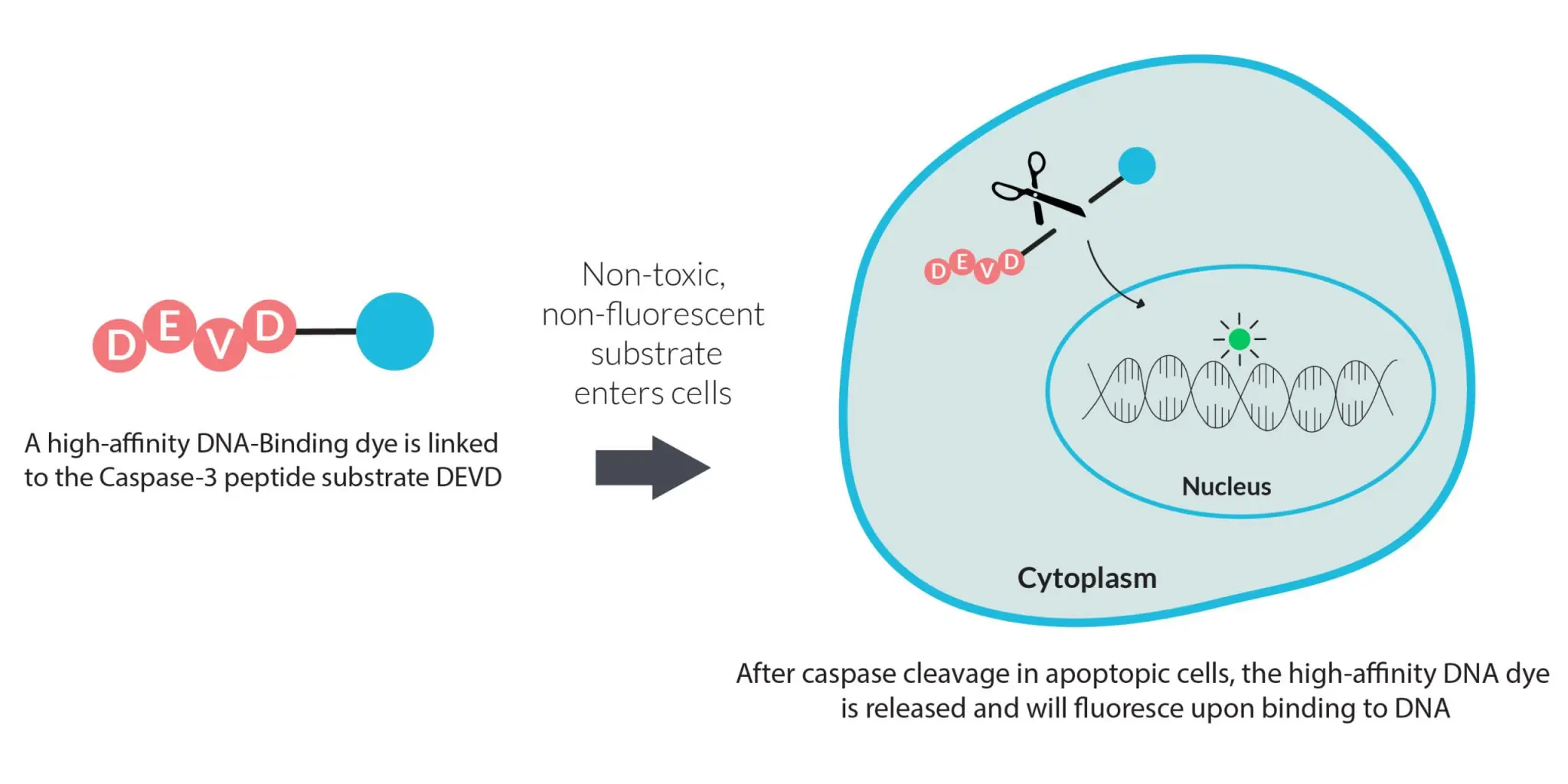
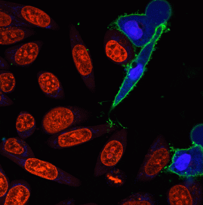
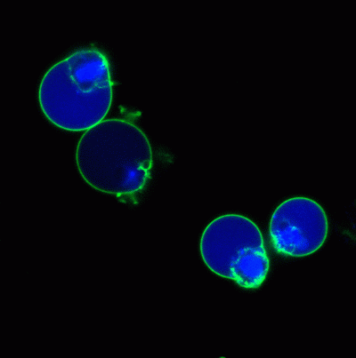
NucView® substrates consist of a fluorogenic DNA dye coupled to the caspase-3/7 DEVD recognition sequence. The substrate, which is initially non-fluorescent, penetrates the plasma membrane and enters the cytoplasm. In apoptotic cells, caspase-3/7 cleaves the substrate, releasing the high-affinity DNA dye, which migrates to the cell nucleus and stains DNA with fluorescence. Thus, NucView caspase-3 substrates are bifunctional, allowing detection of caspase-3/7 activity and visualization of morphological changes in the nucleus during apoptosis. In contrast to other fluorogenic caspase substrates or fluorescent caspase inhibitor based (FLICA) assays, NucView® caspase-3 substrates can be used to detect caspase-3/7 activity within individual intact cells without inhibiting apoptosis progression. Staining is compatible with subsequent fixation and permeabilization for immunostaining.
NucView® 405 dye has excitation/emission at 429/469 nm (with DNA). It is excited by the 405 nm laser line for detection by confocal microscopy in the DAPI channel, or by flow cytometry in the Pacific Blue® channel. NucView® 405 is ideal for caspase-3 detection in multi-color applications for researchers who wish to reserve the green fluorescence channel for other detection reagents.
Note: NucView® 405 signal may be difficult to observe using epifluorescence microscopy. Confocal microscopy using 405 nm laser excitation is recommended for imaging.
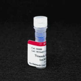
NucView® 405 is also available at 1 mM in PBS (catalog no. 10407), for use in DMSO-sensitive cells.
We also offer green NucView® 488 caspase-3 substrate and kits as well as orange fluorescent NucView® 530.
| Apoptosis/viability marker | Caspase |
|---|---|
| For live or fixed cells | For live/intact cells |
| Assay type/options | Long term staining (24-72h), Endpoint assay, No-wash staining, Real-time imaging, Homogeneous assay |
| Detection method/readout | Fluorescence microscopy, Live cell imaging, Flow cytometry |
| Substrate specificity | Caspases |
| Colors | Blue |
| Fixation options | Fix after staining (formaldehyde), Permeabilize after staining |
| Excitation/Emission | 429/469 nm (with DNA) |
| Storage Conditions | Store at 2 to 8 °C |
Ichimaru, Yoshimi et al.
Sasa veitchii extract induces anticancer effects via inhibition of cyclin D1 expression in MCF-7 cells
Nagoya journal of medical science vol. 82,3 (2020): 509-518.
Article Snippet: “To measure apoptotic cells, we used a Dual Apoptosis Assay Kit with NucView 405 Caspase-3 Substrate (Biotium Inc) and an Annexin V-FITC Detection kit (Nacalai Tesque) according to the manufacturer’s instructions.”
Kim, H., Watanabe, S., Kitamatsu, M. et al.
Cell cycle dependence of apoptosis photo-triggered using peptide-photosensitizer conjugate
Sci Rep 10, 19087 (2020).
DOI: https://doi.org/10.1038/s41598-020-76100-7
Article Snippet: “At 5 h after photoirradiation, the NucView405 Caspase-3 Substrate (Biotium, Fremont, CA, USA) was added to the medium at a final concentration of 100 μM to stain apoptotic cells.”