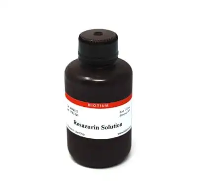
Resazurin Cell Viability Assay offers a simple, rapid, reliable, sensitive, safe and cost-effective measurement of cell viability. This assay has excellent performance compared to other resazurin-based cell proliferation kits such as alamarBlue®, PrestoBlue®, or CellTiter-Blue®.
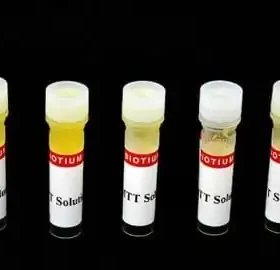
Resazurin detects cell metabolic by converting from a nonfluorescent dye to the highly red fluorescent dye resorufin in response to chemical reduction of growth medium resulting from cell growth. Continued cell growth maintains a reduced environment while inhibition of growth maintains an oxidized environment. Reduction related to growth causes the redox indicator to change from the oxidized form (nonfluorescent, blue) form to the reduced form (fluorescent, red).
Reduced resazurin can be detected using fluorescence (Ex/Em 530-560/590 nm) or absorbance at 570 nm. The fluorescent or colorimetric signal is proportional to the number of living cells in the sample. Results can obtained between 1-24 hours after adding substrate. Resazurin solution is supplied ready-to-use in 25 mL (2500 assay) or 100 mL (10,000 assay) sizes.
See our full selection of Cell Viability & Apoptosis Assays.
| Apoptosis/viability marker | Metabolic activity |
|---|---|
| For live or fixed cells | For live/intact cells |
| Detection method/readout | Microplate reader (absorbance), Microplate reader (fluorescence) |
| Assay type/options | Endpoint assay, Real-time imaging, Homogeneous assay, High-throughput assay |
| Colors | Red |
| Storage Conditions | Store at 2 to 8 °C |
1. Arterioscler, Thromb, Vasc Biol (2010) 30(10), 1940–1948. DOI:10.1161/ATVBAHA.110.205997
2. J Clin Endocrinol Metab, October 2010, 95(10):E181–E191 doi: 10.1210/jc.2010-0581
3. Nucleic Acids Res (2010) 38(22), 8131–8140. doi:10.1093/nar/gkq697
4. Scand J Clin Lab Invest. (2010) 70(7), 512-8. doi: 10.3109/00365513.2010.521255
5. Tissue Eng A (2011) 17(1-2), 37-44. http://doi.org/10.1089/ten.tea.2010.0188
6. Biomaterials (2011) 32(1), 128-36. doi: 10.1016/j.biomaterials.2010.09.006.
7. Food Chem (2012) 135(3), 1533-1538. https://doi.org/10.1016/j.foodchem.2012.06.058
8. Drug Des Dev Therapy (2013) 7, 529–544. http://dx.doi.org/10.2147/DDDT.S45162
9. Langmuir (2013) 29(46), 14254-64. doi: 10.1021/la403533r
10. PLoS ONE (2013) 8(10), e76264. doi:10.1371/journal.pone.0076264
11. ACS Nano (2015) 9(2), 1219-1235. https://doi.org/10.1021/nn504890z
12. Antimicrob Agents Chemother (2016) 60(7), 4183–4196. doi:10.1128/AAC.03021-15
13. Curr Prot Toxicol (2016) 68, 2.24.1-2.24.15. doi: 10.1002/cptx.1
14. J Biol Chem (2016) 291, 21869-21879. doi: 10.1074/jbc.M115.712166
15. Curr Prot Stem Cell Biol (2018) 26(1), 5C.3.1-5C.3.19. DOI:10.1002/9780470151808.sc05c03s26
16. Int J Biol Macromol (2018) 113(1), 132-141. https://doi.org/10.1016/j.ijbiomac.2018.02.069
Contact our Customer Care, Sales & Scientific Assistance
Consult and asked questions about our products & services
Documentation of Technical & Safety Data Sheet, Guides and more...
We gladly support you by keeping you updated on our latest products and the developments around our services.
362 Upper Paya Lebar Rd, #07-15,
Singapore 534963
© 2015-2023 Atlantis Bioscience Pte Ltd. All rights reserved. Co Reg No: 201539516N