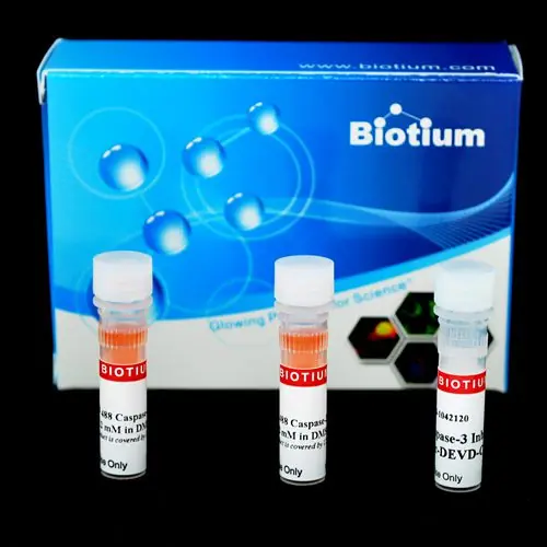
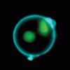
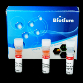
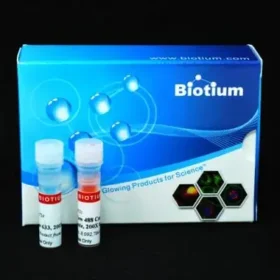
NucView® 488 Caspase-3 Substrate detects apoptosis in intact cells based on caspase-3/7 activity using confocal microscopy, flow cytometry, or live cell imaging.
- NucView® 488 dye has excitation/emission maxima of 500/530 nm (with DNA)
- Rapid, no-wash, endpoint or real-time assays
- Non-toxic, allowing multi-day experiments to be performed
- For flow cytometry, microscopy or live cell imaging systems
- Dual detection of caspase activity and nuclear morphology
- Formaldehyde fixable
NucView® substrates consist of a fluorogenic DNA dye coupled to the caspase-3/7 DEVD recognition sequence. The substrate, which is initially non-fluorescent, penetrates the plasma membrane and enters the cytoplasm. In apoptotic cells, caspase-3/7 cleaves the substrate, releasing the high-affinity DNA dye, which migrates to the cell nucleus and stains DNA with fluorescence. Thus, NucView caspase-3 substrates are bifunctional, allowing detection of caspase-3/7 activity and visualization of morphological changes in the nucleus during apoptosis. In contrast to other fluorogenic caspase substrates or fluorescent caspase inhibitor based (FLICA) assays, NucView® caspase-3 substrates can be used to detect caspase-3/7 activity within individual intact cells without inhibiting apoptosis progression. Staining is compatible with subsequent fixation and permeabilization for immunostaining.
NucView® 488 Substrate in PBS is formulated for use in cells that are sensitive to DMSO toxicity. The substrate also is offered as a 1 mM solution in DMSO (catalog no. 10402). In non-DMSO sensitive cell types, adding DMSO during the substrate incubation may enhance NucView® 488 staining.
We also offer blue fluorescent NucView® 405 Caspase-3 Substrate for use with the 405 nm laser line, and orange fluorescent NucView® 530 for imaging in the Cy®3 channel.
Download a full list of references: Validated cell lines for NucView® 488 with references
Reference Publications
Florian Guisier, Pierre Bohn, et al.
In- and ex-vivo molecular imaging of apoptosis to assess sensitivity of non-small cell lung cancer to EGFR inhibitors using probe-based confocal laser endomicroscopy
PLoS ONE 12(7): e0180576.
DOI: https://doi.org/10.1371/journal.pone.0180576
Article Snippet: “The NucView 488 Caspase-3 substrate (Biotium, San Francisco, California, USA), hereafter referred to as C3-NucView, is a fluorogenic dye that detects caspase 3 activity within intact cells, without interfering with the caspase 3 activity.“
Masahiro YAMAMURA, Akira YAMAUCHI, Shigenobu TONE, Yoshiyuki YAMAGUCHI
Sorafenib-induced apoptosis in colonic neuroendocrine carcinoma cells
Kawasaki Medical Journal 48:175-182,
Article Snippet: “Caspase-3 activation was analysed using the NucView 488 Caspase-3 Assay Kit for live cells (Biotium, San Francisco, CA, USA), a caspase-3 substrate that fluoresces when cleaved.”
Vasconcelos et al.
Individual Human Cytotoxic T Lymphocytes Exhibit Intraclonal Heterogeneity during Sustained Killing
Cell Reports 11, 1474–1485
DOI: http://dx.doi.org/10.1016/j.celrep.2015.05.002
Article Snippet: “For live cell imaging, 1 3 105 pulsed target cells were seeded into microchambers (Lab-Tek Chambered coverglass; Nalgen Nunc) previously coated with poly-D lysine, followed by addition of 2 mM NucView 488 caspase-3 substrate (Biotium)”
Maurice Bosch and Vernonica E. Franklin-Tong
Temporal and spatial activation of caspase-like enzymes induced by self-incompatibility in Papaver pollen
PNAS 104 (46) 18327-18332
DOI: https://doi.org/10.1073/pnas.0705826104
Article Snippet: “The NucView 488 caspase-3 assay Kit (Biotium, Inc., Hayward, CA) was used to detect DEVDase activity in live pollen tubes. The specificity of the DEVDase activity was checked by pretreating hydrated pollen for 1 h with 100 μM Ac-DEVD-CHO or Ac-YVAD-CHO before SI induction.”