Partnering with Biowest, let’s unlock precise, gentle, and reliable cell detachment with our Trypsin 0.25% – EDTA formulation, designed to meet the highest standards in cell culture workflows. Optimized with a balanced HBSS buffer (free of calcium and magnesium) and enhanced with phenol red for easy pH monitoring, this solution ensures minimal cell stress and maximum viability during passaging or tissue dissociation.
Derived from porcine pancreas, our high-quality trypsin is paired with EDTA to disrupt cell–cell adhesion, making it ideal for a wide range of mammalian cells. Stringently filtered for sterility and stability, this ready-to-use reagent guarantees consistency, offering confidence with every use.
Optimal Enzyme Concentration
Contains 0.25% trypsin, delivering efficient cell detachment while preserving cell integrity.
EDTA-Enhanced Performance
EDTA chelates divalent cations, promoting rapid and gentle dissociation of even tightly adherent cells.
HBSS Buffer Base
Formulated without calcium and magnesium to enhance dissociation efficiency and reduce re-aggregation.
Phenol Red Indicator
Enables real-time pH monitoring to maintain optimal physiological conditions during handling.
Strict Sterility and Stability
Sterile-filtered and supplied frozen for long-term reliability (24-month shelf life when stored at -18°C to -40°C).
Versatile Application
Ideal for routine cell culture maintenance, subculturing, and primary cell isolation.
Trust Biowest for your cell culture essentials.
Please enquire with us at [email protected]
We deliver Biowest solutions across South East Asia (Singapore, Thailand, Vietnam, Indonesia, Philippines, Myanmar) and Across China Only.
| Attribute | Details |
|---|---|
| Product Code | L0931-100 |
| State | Frozen |
| Shipping | Gel pack, dry ice, or refrigerated |
| Shelf Life | 24 months |
| Storage | -18°C to -40°C |
| Applications | Cell Culture, Tissue Dissociation |
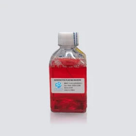
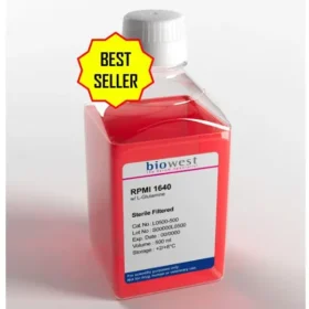
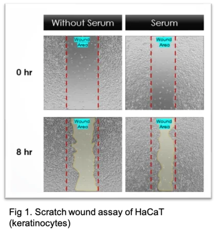
Biowest Trypsin 0.25 % in PBS w/o Calcium w/o Magnesium w/o Phenol Red (L0931-100 ) Used For Scratch Wound Assay
Wound healing assays (Scratch wound assay) are widely utilized in cancer research and studies of cell-cell interactions to investigate cell migration, particularly in the context of skin wound repair and cancer metastasis. In this method, a confluent monolayer of cells is mechanically scratched to create a “wound,” and the rate of closure is monitored over time.
In the experiment, HaCaT were routinely cultured Dulbecco’s modified Eagle’s medium (DMEM) supplemented with 10% fetal bovine serum (FBS). All cells were cultured in a 37 degree Celsius, 5% carbon dioxide, humidified incubator. keratinocytes were routinely sub-cultured using 0.25 % trypsin and 1 mM Ethylenediaminetetraacetic acid. A linear scratch wound were created on a confluent layer of seeded cells and were treated in DMEM low glucose in the absence or presence of 10% FBS for 8 hours. Images were captured at regular intervals to analyse the wound closure over time.
Materials:
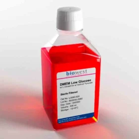
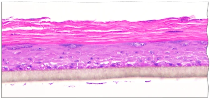
Biowest Trypsin 0.25 % in PBS w/o Calcium w/o Magnesium w/o Phenol Red (L0931-100 ) Used For 3D In-vitro Skin
The EU has banned animal testing for cosmetics and their ingredients since 2013. A growing demand for in vitro skin models has been rising steadily to cater for both the safety testing (Skin irritation) and efficacy testing (functional test).
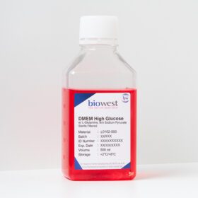
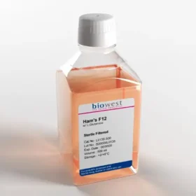
In the experiment, primary keratinocytes were seeded on a cultured insert to form a confluent layer. The confluent layer of cells is lifted to an air-liquid interface for 14 days to stimulates the keratinocytes to differentiate (stratification) and form a multi-layered epidermis that contains stratum corneum layers. The specialized medium primarily used consists of DMEM and Ham’s F12 in a specific ratio. It is chemically defined and serum-free to ensure a high reproducibility in result. Treatments can be topically applied to the skin model for a period of times. Histology such as H&E staining and other skin markers are commonly carried out. Gene expression and protein study are also alternative analysis method.
Materials:
Contact our Customer Care, Sales & Scientific Assistance
Consult and asked questions about our products & services
Documentation of Technical & Safety Data Sheet, Guides and more...
362 Upper Paya Lebar Rd, #07-15,
Singapore 534963
© 2015-2023 Atlantis Bioscience Pte Ltd. All rights reserved. Co Reg No: 201539516N