A red fluorescent dead cell stain for bacteria and mammalian cells
| SIZE |
CATALOG #
|
PRICE
|
| 1 mL | #40050 |
$250 |
| Cellular localization |
Nucleus & cytoplasm |
|---|---|
| Assay type/options |
Real-time imaging |
| Cell permeability |
Membrane impermeant |
| Apoptosis/viability marker |
Dead cell stain |
| Colors |
Red |
| Excitation/Emission |
279, 522/593 nm (with DNA) |
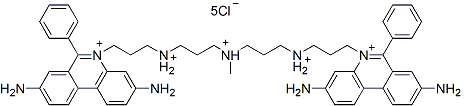
Ethidium Homodimer III, also known as EthD-III, is a red fluorescent dead cell stain for bacteria and mammalian cells. It is a cell membrane-impermeant nucleic acid dye that stains only dead cells with damaged cell membranes. Ethidium Homodimer III was developed by Biotium as a superior alternative to Ethidium Homodimer I. The absorption and emission spectra are similar, but EthD-III is 45% brighter.
Ethidium Homodimer III is membrane impermeant and useful for selectively staining dead cells with damaged plasma membranes. It also is a component of several combination viability assay kits for detecting both live and dead cells in the same population, such as our Viability/Cytotoxicity Assay Kit for Animal Cells (Cat. No 30002) and our Viability/Cytotoxicity Assay Kit for Bacteria (Cat. No. 30027).
We also offer Ethidium Homodimer III, 1 mM solution in DMSO (catalog no. 40051).
EthD-III is dead cell specific in all cell types, including mammalian cells, bacteria and yeast. See our Cellular Stains Table for more information on how our dyes stain various organisms.
Yan, Wei-Tao et al. PANoptosis-like cell death in ischemia/reperfusion injury of retinal neurons Neural regeneration research vol. 18,2 (2023): 357-363. DOI: 10.4103/1673-5374.346545 Article Snippet: “Ethidium Homodimer III (EthD-III; Biotium, Fremont, CA, USA, Cat# 40050) staining combined with DSF was used to indicate pyroptosis. DSF inhibits GSDMD expression and interferes with pore formation triggered by GSDMD in the cell membrane to prevent the release of interleukin and other inflammatory factors (Zhang et al., 2021a, b).” Hsieh, Huai-Ching et al. Characterization and identification of cell death dynamics by quantitative phase imaging Journal of biomedical optics vol. 27,4 (2022): 046502. DOI: 10.1117/1.JBO.27.4.046502 Article Snippet: “After treating the cells for 1.5 h, we stained the cells by CellEvent Caspase-3/7 Green Detection Reagent (Caspase-3/7, ThermoFisher Scientific) and Ethidium Homodimer III (EthD-III, Biotium), a DNA dye for dead cells only.” Dong, Y., Huang, Y., Zhang, Z. et al. iRGD-modified memory-like NK cells exhibit potent responses to hepatocellular carcinoma J Transl Med 21, 205 (2023). DOI: https://doi.org/10.1186/s12967-023-04024-7 Article Snippet: “After incubation, MCSs were treated with Calcein AM and Ethidium homodimer III (EthD-III) solutions (Viability/Cytotoxicity Assay Kit, biotium, USA) as per the manufacturer’s instructions.” Tyszka-Czochara M, Konieczny P, Majka M. Caffeic Acid Expands Anti-Tumor Effect of Metformin in Human Metastatic Cervical Carcinoma HTB-34 Cells: Implications of AMPK Activation and Impairment of Fatty Acids De Novo Biosynthesis International Journal of Molecular Sciences. 2017; 18(2):462. DOI: https://doi.org/10.3390/ijms18020462 Article Snippet: “Fluorescent dyes Annexin-V (excitation/emission 490/515 nm) and Ethidium homodimer (EthD-III, excitation/emission 528/617 nm) were used (Biotium, Fremont, CA, USA). The cells were gated according to forward (FSC), side scatter (SSC), and appropriate fluorescence parameters.” Maeda, E., Ando, Y., Takeshita, K. et al. Through the cleared aorta: three-dimensional characterization of mechanical behaviors of rat thoracic aorta under intraluminal pressurization using optical clearing method Sci Rep 12, 8632 (2022). DOI: https://doi.org/10.1038/s41598-022-12429-5 Article Snippet: “The specimen was incubated in 5 µM ethidium homodimer-III (Biotium, USA) in PBS overnight at 4 °C to stain the nuclei of smooth muscle cells and was imaged under a two-photon microscope (A1R MP, Nikon, Japan) in a noncleared, normal state within PBS.”

Contact our Customer Care, Sales & Scientific Assistance
Consult and asked questions about our products & services
Documentation of Technical & Safety Data Sheet, Guides and more...
We gladly support you by keeping you updated on our latest products and the developments around our services.
362 Upper Paya Lebar Rd, #07-15,
Singapore 534963
© 2015-2023 Atlantis Bioscience Pte Ltd. All rights reserved. Co Reg No: 201539516N