
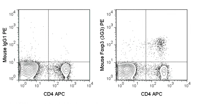
The 3G3 antibody reacts with mouse Foxp3, a 50-55 kDa transcription factor which is a central regulator of T cell activity and is critical for the development and function of regulatory T cells (Tregs). Foxp3 is expressed at constitutively high levels in Treg cells, which are further identified as being CD4+ CD25+. In resting conventional T cells (CD4+ CD25-) Foxp3 expression is restricted, and upon TCR activation is expressed only transiently and in a small proportion of cells. However, the growth factor TGF-beta has been shown to induce expression of Foxp3 in naïve T cells, driving their development into Foxp3+ Tregs, which are called “induced” or “adaptive” Tregs. These cells are phenotypically similar to so-called “natural” Tregs (CD4+ CD25high Foxp3+) which originate in the thymus and comprise the majority of Treg cells. Tregs are critical for maintaining peripheral tolerance and are implicated in the development of autoimmunity.
It is important to review the literature in choosing an antibody for the Foxp3 antigen in flow cytometry, as the potential for high background or non-specific staining may be observed. The 3G3 antibody may be used for intracellular detection of Foxp3 in cells from mouse and Rhesus macaque.
| Name | PE Anti-Mouse Foxp3 (3G3) |
|---|---|
| Cat. No. | 50-5773 |
| Alternative Names | Forkhead Box P3, Scurfin |
| Gene ID | 50943/20371 |
| Clone | 3G3 |
| Isotype | Mouse IgG1, kappa |
| Reactivity | Mouse |
| Cross Reactivity | Rhesus macaque |
| Format | PE |
| Application | Flow Cytometry |
| Citations* | Ramos RN, Oliveira CE, Gasparoto TH, et al. 2012. Carcinogenesis. 33: 902-909. (Flow cytometry)
Klein M, Vaeth M, Scheel T, Grabbe S, Baumgrass R, Berberich-Siebelt F, Bopp T, Schmitt E, and Becker C. 2012. J. Immunol. 188: 1091-1097. (Flow cytometry) Ansari AA, Reimann KA, Mayne AE, Takahashi Y, Stephenson ST, Wang R, Wang X, Li J, Price AA, Little DM, Zaidi M, Lyles R, and Villinger F. 2011. J. Immunol. 186: 1044-1059. (Flow cytometry – Rhesus macaque) Nagar M, Vernitsky H, Cohen Y, Dominissini D, Berkun Y, Rechavi G, Amariglio N, and Goldstein I. 2008. Int. Immunol. 20: 1041-1055. (Flow cytometry) Hombach AA, Kofler D, Hombach A, Rappl G, and Abken H. 2007. J. Immunol. 179: 7924-7931. (Flow cytometry). Gavin MA, Torgerson TR, Houston E, deRoos P, Ho WY, Stray-Pedersen A, Ocheltree EL, Greenberg PD, Ochs HD, and Rudensky AY. 2006. Proc Natl Acad Sci USA. 103(17): 6659-6664. (Flow cytometry) |
Application Key:FC = Flow Cytometry; FA = Functional Assays; ELISA = Enzyme-Linked Immunosorbent Assay; ICC = Immunocytochemistry; IF = Immunofluorescence Microscopy; IHC = Immunohistochemistry; IHC-F = Immunohistochemistry, Frozen Tissue; IHC-P = Immunohistochemistry, Paraffin-Embedded Tissue; IP = Immunoprecipitation; WB = Western Blot; EM = Electron Microscopy
*Tonbo Biosciences tests all antibodies by flow cytometry. Citations are provided as a resource for additional applications that have not been validated by Tonbo Biosciences. Please choose the appropriate format for each application and consult the Materials and Methods section for additional details about the use of any product in these publications.


Contact our Customer Care, Sales & Scientific Assistance
Consult and asked questions about our products & services
Documentation of Technical & Safety Data Sheet, Guides and more...
We gladly support you by keeping you updated on our latest products and the developments around our services.
362 Upper Paya Lebar Rd, #07-15,
Singapore 534963
© 2015-2023 Atlantis Bioscience Pte Ltd. All rights reserved. Co Reg No: 201539516N


