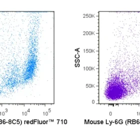
The RB6-8C5 antibody binds to mouse Ly-6G, commonly known as Gr-1, a member of the Ly-6 superfamily of GPI-anchored cell surface proteins with roles in cell signaling and cell adhesion. Gr-1 is differentially expressed during development and maturation of cells in the myeloid lineage and is expression at varying stages and levels on monocytes, macrophages, granulocytes, and peripheral neutrophils. In the mouse, the RB6-8C5 antibody is typically used in combination with the macrophage labeling antibody M1/70 (Anti-CD11b) for phenotypic analysis of monocytes, macrophages and granulocytes.
Note: The RB6-8C5 antibody has been reported to cross-react with Ly-6C on cells expressing this antigen (Fleming et al. 1993. J. Immunol. 151:2399-2408 and Sasmono et al. 2007. J. Leukoc. Biol. 82: 111-123) and has been cited in the literature for identification of Ly-6G/Ly-6C. Other reports suggest that this antibody is specific for Ly-6G, without cross-reactivity for Ly-6C (Nagendra S. and Schlueter AJ. 2003. Cytometry A, 58(2): 195-200.
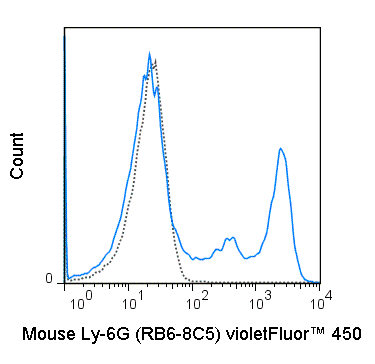
| Name | violetFluor™ 450 Anti-Mouse Ly-6G (Gr-1) (RB6-8C5) |
|---|---|
| Cat. No. | 75-5931 |
| Alternative Names | Gr1, Ly6G |
| Gene ID | |
| Clone | RB6-8C5 |
| Isotype | Rat IgG2b, κ |
| Reactivity | Mouse |
| Cross Reactivity | |
| Format | violetFluor™ 450 |
| Application | Flow Cytometry |
| Citations* | Berent-Maoz B, Montecino-Rodriguez E, Signer RAJ, and Dorshkind K. 2012. Blood. 199:5715-5721. (Flow cytometry)
von Bruhl M-L, Stark K, Steinhart A, et al. 2012. J. Exp. Med. 209: 819-835. (Intravital fluorescent microscopy – video) Le HT, Tran VG, Kim W, Kim H, Cho HR, and Kwon B. 2012. J. Immunol. 189:287-295. (in vivo neutrophil depletion) Doring Y, Soehnlein O, Drechsler M, Shagdarsuren E, Chaudhari SM, Meiler S, Hartwig H, Hristov M, Koenen RR, Hieronymus T, Zenke M, Weber C, and Zernecke A. 2012. Arterioscler. Thromb. Vasc. Biol. 32: 1613-1623. (in vivo depletion) Hickman HD, Li L, Reynoso GV, Rubin EJ, Skon CN, Mays JW, Gibbs J, Schwartz O, Bennink JR, and Yewdell JW. 2011. J. Exp. Med. 208: 2511-2524. (Immunohistochemistry – OCT embedded frozen tissue) Wang T, Tian L, Haino M, Gao J-L, Lake R, Ward Y, Wang H, Siebenlist U, Murphy PM, and Kelly K. 2007. Infect. Immun. 75(3):1144-1153. (Immunohistochemistry – zinc fixed tissue) Nutt SL, Metcalf D, D’Amico A, Polli M, and Wu L. 2005. J. Exp. Med. 201:221-231. (Immunomagnetic bead depletion) Whiteland JL, Nicholls SM, Shimeld C, Easty DL, Williams NA, and Hill TJ. 1995. J. Histochem. Cytochem. 43:313-320. (Immunohistochemistry – frozen tissue, paraffin embedded tissue) Fleming TJ, Fleming ML, and Malek TR. 1993. J. Immunol. 151:2399-2408. (in vitro blocking, immunoprecipitation) |
Application Key:FC = Flow Cytometry; FA = Functional Assays; ELISA = Enzyme-Linked Immunosorbent Assay; ICC = Immunocytochemistry; IF = Immunofluorescence Microscopy; IHC = Immunohistochemistry; IHC-F = Immunohistochemistry, Frozen Tissue; IHC-P = Immunohistochemistry, Paraffin-Embedded Tissue; IP = Immunoprecipitation; WB = Western Blot; EM = Electron Microscopy
*Tonbo Biosciences tests all antibodies by flow cytometry. Citations are provided as a resource for additional applications that have not been validated by Tonbo Biosciences. Please choose the appropriate format for each application and consult the Materials and Methods section for additional details about the use of any product in these publications.


Contact our Customer Care, Sales & Scientific Assistance
Consult and asked questions about our products & services
Documentation of Technical & Safety Data Sheet, Guides and more...
We gladly support you by keeping you updated on our latest products and the developments around our services.
362 Upper Paya Lebar Rd, #07-15,
Singapore 534963
© 2015-2023 Atlantis Bioscience Pte Ltd. All rights reserved. Co Reg No: 201539516N