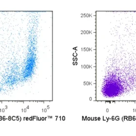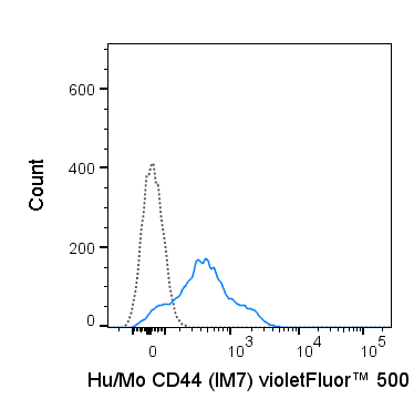
The IM7 antibody recognizes CD44, a ubiquitously expressed cell surface receptor which is important for extracellular matrix organization, cell-cell and cell-matrix adhesion and migration. CD44 may be expressed in a number of different isoforms (splice variants) from the most typical or “standard” form, known as CD44s, to variants designated CD44v, e.g. CD44v1 or CD44v6. These receptors interact with several ligands, but most often associate with an extracellular matrix component hyaluronate, through which it mediates adhesion.
The IM7 antibody may be used for detection of all isoforms of CD44, as it recognizes constant epitopes near the extracellular proximal domain. (Xu et al, 2002, J. Leukoc. Biol. 72:1133-1141). It has been reported to be cross-reactive with many non-human species including Baboon, Chimpanzee, Cynomolgus, Rhesus, Horse, Cow, Pig, Dog and Cat CD44.
| Name | violetFluor™ 500 Anti-Human/Mouse CD44 (IM7) |
|---|---|
| Cat. No. | 85-0441 |
| Alternative Names | Pgp-1, MDU3, Hermes, Hyaluronate receptor |
| Gene ID | 12505 |
| Clone | IM7 |
| Isotype | Rat IgG2b, κ |
| Reactivity | Human, Mouse |
| Cross Reactivity | Baboon, Canine, Chimpanzee, Cynomolgus, Equine, Feline, Rhesus, Swine |
| Format | violetFluor™ 5000 |
| Application | Flow Cytometry |
| Citations* | Chandler HL, Haeussler DJ, Gemensky-Metzler AJ, Wilkie DA, and Lutz EA. 2012. Invest. Ophthalmol. Vis. Sci. 53:1835-1845. (in vitro blocking, canine)
Lee L-F, Logronio K, Tu GH, Zhai W, Ni I, Mei L, Dilley J, Yu J, et al. 2012. Proc. Natl. Acad. Sci. 10.1073. (Flow cytometry). Ruffell B, Poon GFT, Lee SSM, Brown KL, Tjew S-L, Cooper J, and Johnson P. 2011. J. Biol. Chem. 286:19179-19190. (Immunoprecipitation) Miyake Y, Matsumoto H, Yokoo M, Miyazawa K et al. 2006. Biol. Reprod. 74: 501-510. (Immunohistochemistry – frozen tissue, swine) Veir JK, Lappin MR, and Dow SW. 2006. Journal of Feline Medicine and Surgery. 8:400-411. (Flow cytometry – feline) Frank NY, Margaryan A, Huang Y, Schatton T, Waaga-Gasser AM, Gasser M, Sayegh MH, Sadee W, and Frank MH. 2005. Cancer Res. 65:4320-4333. (Immunohistochemistry – frozen tissue) Fischer A, Schumacher N, Maier M, Sendtner M, and Gessler M. 2004. Genes & Dev. 18:901-911. (Immunohistochemistry – paraffin embedded tissue) Xu H, Manivannan A, Liversidge J, Sharp PF, Forrester JV, and Crane IJ. 2002. J. Leukoc. Biol. 72:1133-1141. (in vivo functional assays, induction of apoptosis) Si-Tahar M, Sitaraman S, Shibahara T, and Madara JL. 2001. Am. J. Physiol. Cell Physiol. 280:C423-C432. (in vitro functional assays, Western Blot) |
Application Key:FC = Flow Cytometry; FA = Functional Assays; ELISA = Enzyme-Linked Immunosorbent Assay; ICC = Immunocytochemistry; IF = Immunofluorescence Microscopy; IHC = Immunohistochemistry; IHC-F = Immunohistochemistry, Frozen Tissue; IHC-P = Immunohistochemistry, Paraffin-Embedded Tissue; IP = Immunoprecipitation; WB = Western Blot; EM = Electron Microscopy
*Tonbo Biosciences tests all antibodies by flow cytometry. Citations are provided as a resource for additional applications that have not been validated by Tonbo Biosciences. Please choose the appropriate format for each application and consult the Materials and Methods section for additional details about the use of any product in these publications.


Contact our Customer Care, Sales & Scientific Assistance
Consult and asked questions about our products & services
Documentation of Technical & Safety Data Sheet, Guides and more...
We gladly support you by keeping you updated on our latest products and the developments around our services.
362 Upper Paya Lebar Rd, #07-15,
Singapore 534963
© 2015-2023 Atlantis Bioscience Pte Ltd. All rights reserved. Co Reg No: 201539516N