PE Anti-Mouse CD357 (AITR / GITR) (DTA-1)

PE Anti-Mouse CD357 (AITR / GITR) (DTA-1)

PE Anti-Mouse CD357 (AITR / GITR) (DTA-1)

Add to cart

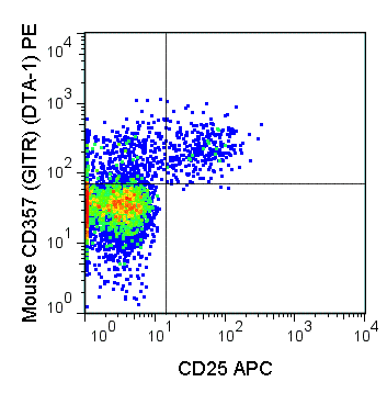
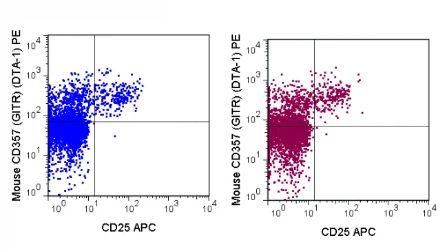
The DTA-1 antibody reacts with mouse CD357, also known as GITR or AITR (in humans), a 66-70 kDa member of the Tumor Necrosis Factor superfamily (TNFRSF18). GITR is primarily found on T cells, and its function may vary depending on the T cell type where it is expressed. GITR is upregulated on activated T cells where it provides co-stimulation, yet GITR may promote the inhibition of CD4+ CD25+ Treg cells, where it is expressed at high levels. GITR ligand (GITRL) is found on B cells, macrophages, dendritic and endothelial cells, and is implicated in regulating both innate and adaptive immune responses.

The DTA-1 antibody may be used for analysis of GITR expression on T cells, and is also commonly used in vitro as an agonistic antibody to induce GITR signaling in various assays (use format appropriate for functional assays).

Product Details

Name PE Anti-Mouse CD357 (GITR) (DTA-1)
Cat. No. 50-5874
Alternative Names TNFRSF18
Gene ID 21936
Clone DTA-1
Isotype Rat IgG2b
Reactivity Mouse
Cross Reactivity
Format PE
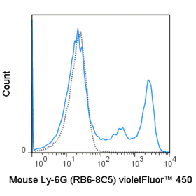
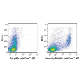
Application Flow Cytometry
Citations* Lee L-F, Logronio K, Tu GH, Zhai W, Ni I, Mei L, Dilley J, Yu J, et al. 2012. Proc. Natl. Acad. Sci. 10.1073. (Flow cytometry).

Joetham A, Ohnishi H, Okamoto M, Takeda K, Schedel M, Domenico J, Dakhama A, and Gelfand EW. 2012. J. Biol. Chem. 287: 17100-17108. (in vitro activation)

Van der Werf N, Redpath SA, Phythian-Adams AT, Azuma M, Allen JE, Maizels RM, Macdonald AS, and Taylor MD. 2011. J. Immunol. 187: 1411-1420. (in vivo activation)

Molloy MJ, Zhang W, and Usherwood EJ. 2011. J. Immunol. 186: 6218-6226. (in vivo activation) ll

Yokoyama T, Matsuda S, Takae Y, Wada N, Nishikawa T, Amagai M, and Koyasu S. 2011. Int. Immunol. 23: 365-373. (Treg depletion – magnetic beads)

Application Key:FC = Flow Cytometry; FA = Functional Assays; ELISA = Enzyme-Linked Immunosorbent Assay; ICC = Immunocytochemistry; IF = Immunofluorescence Microscopy; IHC = Immunohistochemistry; IHC-F = Immunohistochemistry, Frozen Tissue; IHC-P = Immunohistochemistry, Paraffin-Embedded Tissue; IP = Immunoprecipitation; WB = Western Blot; EM = Electron Microscopy

*Tonbo Biosciences tests all antibodies by flow cytometry. Citations are provided as a resource for additional applications that have not been validated by Tonbo Biosciences. Please choose the appropriate format for each application and consult the Materials and Methods section for additional details about the use of any product in these publications.

Technical Data Sheet                      View All Formats                  Request Custom/Bulk