
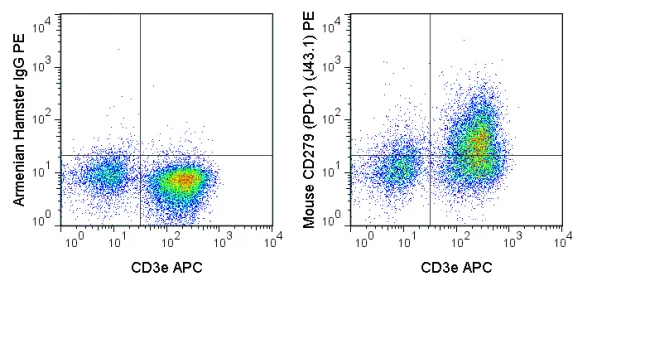
The J43.1 antibody is specific for mouse CD279, also known as programmed death-1 (PD-1), a 55 kDa glycoprotein which can co-regulate T cell antigen receptor signaling and therefore modulate T cell activation. PD-1 exists in a monomeric form that is expressed by CD4- CD8- thymocytes, where it participates in the processes of clonal selection, elimination of autoreactive lymphocytes, and development of tolerance. PD-1 expression is also inducible upon activation of mature T cells, where it has been proposed to interact with the co-stimulatory receptor CD80 to limit T cell activation. Two ligands for PD-1, known as PD-L1 (B7-H1) and PD-L2 (B7-DC) are differentially expressed on T and B cells, monocytes, macrophages, NK cells or dendritic cells. PD-1 is a member of a family of receptors including CD28 and CTLA-4 (CD152), which interact with “B7” ligands to provide a balance of co-stimulatory /co-inhibitory signaling important in T cell activation, tolerance, and autoimmunity.
The J43.1 antibody may be used as a marker for PD-1 expression, and is commonly used for analysis of receptor-ligand interaction and function(s) in vitro and in vivo. Please choose the appropriate format for each application.
| Name | PE Anti-Mouse CD279 (PD-1) (J43.1) |
|---|---|
| Cat. No. | 50-9985 |
| Alternative Names | PDCD1, PD1, SLEB2 , PD-1 |
| Gene ID | 18566 |
| Clone | J43.1 |
| Isotype | Armenian Hamster IgG |
| Reactivity | Mouse |
| Cross Reactivity | |
| Format | PE |
| Application | Flow Cytometry |
| Citations* | Grinberg-Bleyer Y, Oh H, Desrichard A, Bhatt DM, Caron R, Chan TA, Schmid RM, Klein U, Hayden MS and Ghosh S. 2017. NF-κB c-Rel Is Crucial for the Regulatory T Cell Immune Checkpoint in Cancer. Cell. doi: 10.1016/j.cell.2017.08.004.
Hams E, McCarron MJ, Amu S, Yagita H, Azuma M, Chen L, and Fallon PG. 2011. J. Immunol. 186:5648-5655. (in vivo blocking) Rivas MN, Weatherly K, Hazzan M, Vokaer B, Dremier S, Gaudray F, Goldman M, Salmon I, and Braun MY. 2009. 183:4284-4291. (in vitro blocking) Koehn BH, Ford ML, Ferrer IR, Borom K, Gangappa S, Kirk AD, and Larsen CP. 2008. J. Immunol. 181:5313-5322. (in vivo blocking) Brooks DG, Ha S-J, Elsaesser H, Sharpe AH, Freeman GJ, and Oldstone MBA. 2008. Proc. Natl. Acad. Sci. 105:20428-20433. (Flow cytometry) Ansari MJI, Salama AD, Chitnis T, Smith RN, Yagita H, Akiba H, Yamazaki T, Azuma M, Isai H, Khoury SJ, Auchincloss H, and Sayegh MH. 2003. J. Exp. Med. 198:63-71. (Immunohistochemistry – frozen tissue, in vivo blocking) Agata Y, Kawasaki A, Nishimura H, Ishida Y, Tsubat T, Yagita H, and Honjo T. 1996. Int. Immunol. 8:765-772. (Immunoprecipitation) |
Application Key:FC = Flow Cytometry; FA = Functional Assays; ELISA = Enzyme-Linked Immunosorbent Assay; ICC = Immunocytochemistry; IF = Immunofluorescence Microscopy; IHC = Immunohistochemistry; IHC-F = Immunohistochemistry, Frozen Tissue; IHC-P = Immunohistochemistry, Paraffin-Embedded Tissue; IP = Immunoprecipitation; WB = Western Blot; EM = Electron Microscopy
*Tonbo Biosciences tests all antibodies by flow cytometry. Citations are provided as a resource for additional applications that have not been validated by Tonbo Biosciences. Please choose the appropriate format for each application and consult the Materials and Methods section for additional details about the use of any product in these publications.


Contact our Customer Care, Sales & Scientific Assistance
Consult and asked questions about our products & services
Documentation of Technical & Safety Data Sheet, Guides and more...
We gladly support you by keeping you updated on our latest products and the developments around our services.
362 Upper Paya Lebar Rd, #07-15,
Singapore 534963
© 2015-2023 Atlantis Bioscience Pte Ltd. All rights reserved. Co Reg No: 201539516N


