LysoView™ Dyes

LysoView™ Dyes

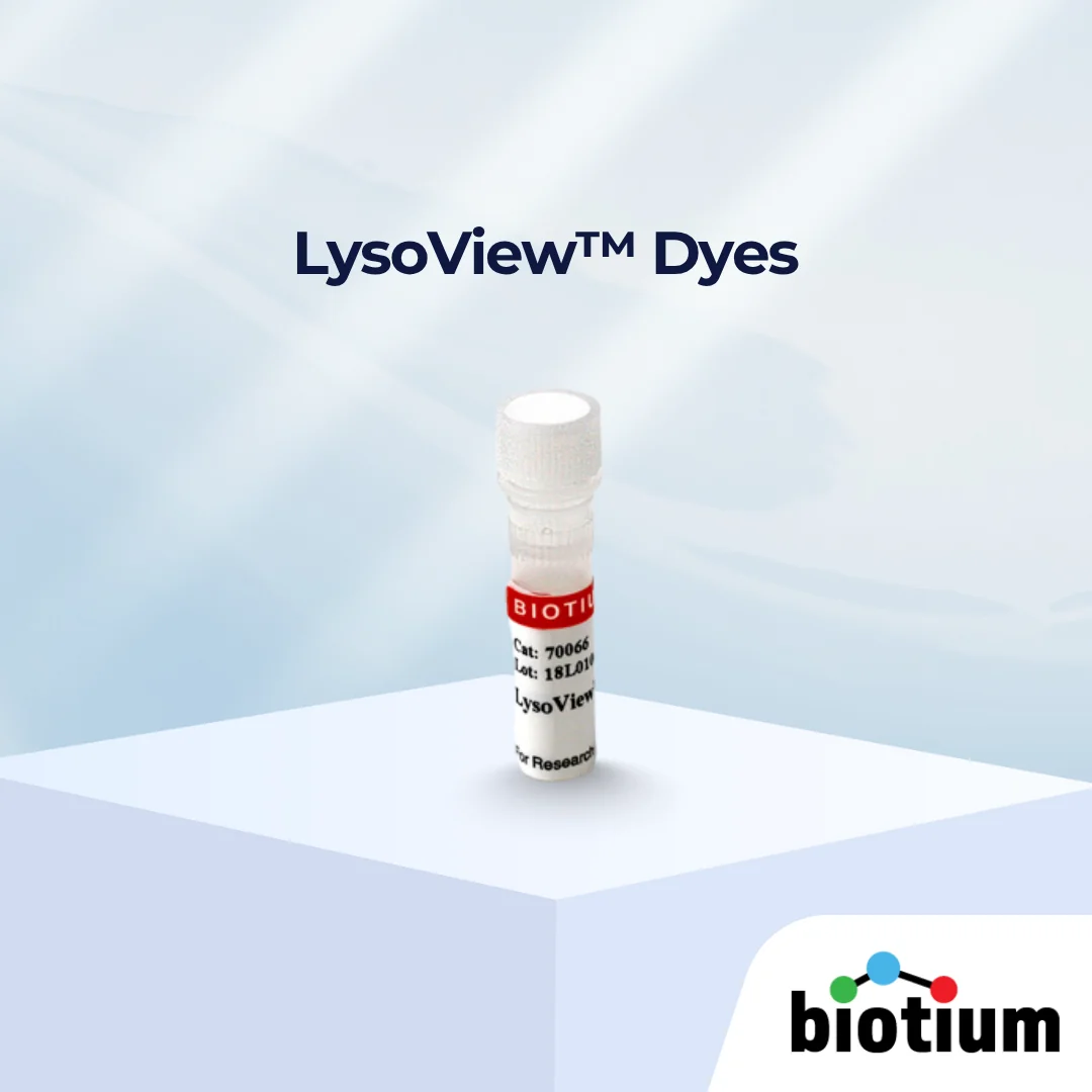
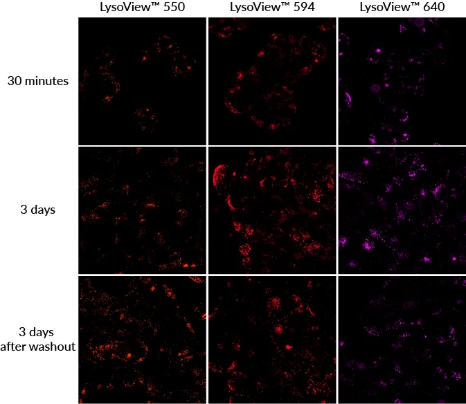
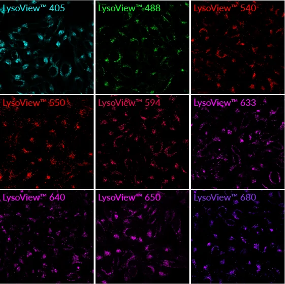
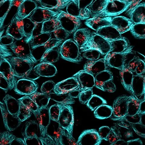
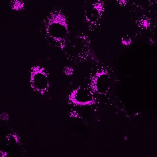
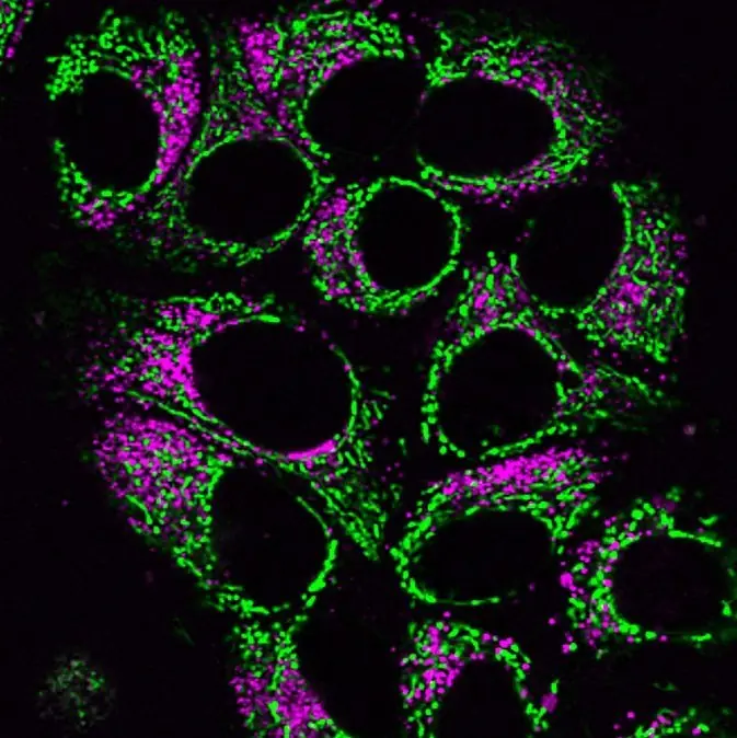
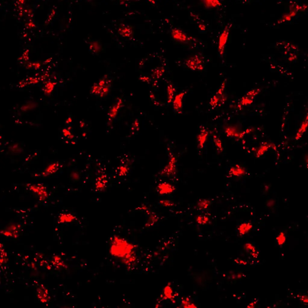
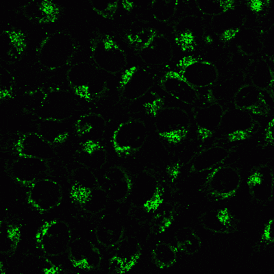
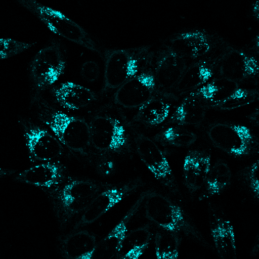
Bright no-wash fluorescent dyes for live-cell lysosome imaging. Designed for high specificity, low background and long-term imaging, with colours from blue to near-IR and options validated for super-resolution microscopy. Ideal for studying lysosomal function, autophagy and cell health.

LysoView™ Dyes cover image

Add to cart
SKU: 70066-T, 70066, 70067-T, 70067, 70061-T, 70061, 70083-T, 70083, 70084-T, 70084, 70058-T, 70058, 70085-T, 70085, 70059-T, 70059, 70086-T, 70086 Categories: Brand:

LysoView™ Dyes – No-Wash Lysosomal Stains for Live-Cell Imaging

LysoView™ dyes are advanced fluorescent probes designed for high-specificity lysosome staining in live cells, offering bright, low-background fluorescence without a wash step. Available in a full spectrum from blue to near-infrared, LysoView™ provides flexible options for confocal, SIM, STED and long-term live-cell imaging.

Perfect for researchers studying lysosomal biology, autophagy, cell stress, neurodegeneration and drug response.

Key Benefits

  • No-wash lysosome staining for clean, high-contrast images
  • Non-toxic formulation supports real-time and long-term imaging (24–72 h)
  • Brighter and lower background compared to conventional lysosomal probes
  • Validated for super-resolution: LysoView™ 488 (SIM), LysoView™ 650 (SIM and STED)
  • pH-sensitive options (LysoView™ 540, LysoView™ 633) for enhanced specificity
  • Wide colour range: blue, green, orange, red, far-red and unique NIR
  • Flexible trial sizes for convenient assay testing
  • Cost-efficient per assay with concentrated 1000X format


Product Features

  • Targets lysosomes in live/intact cells
  • Membrane-permeant fluorescence for immediate staining
  • Compatible with super-resolution microscopy
  • Photostable dyes (550, 594, 640, 650) ideal for prolonged imaging
  • Storage at −10 to −35 °C, light-protected
  • Available in 10 µL trials, 50 µL vials and lyophilised formats


Applications

  • Lysosome localisation and morphology studies
  • Autophagy and endolysosomal dynamics
  • Drug-induced lysosomal perturbation
  • High-content screening
  • Super-resolution lysosomal mapping

Specification

Attribute Specification
Size Trial size (10 µL), 50 µL, Trial size (1 vial), Set of 10 vials
Probe cellular localisation Lysosomes
Dye LysoView™ 405, LysoView™ 488, LysoView™ 540, LysoView™ 550, LysoView™ 594, LysoView™ 633, LysoView™ 640, LysoView™ 650, LysoView™ 680
For live or fixed cells For live/intact cells
Assay type/options Long-term staining (24–72 h), No-wash staining, Real-time imaging
Cell permeability Membrane permeant
Colours Blue, Green, Orange, Red, Far-red, Near-infrared
Storage conditions Store at −10 to −35 °C, Protect from light

Variations

Product Catalogue Number Size Abs/Em (nm) pH ≤ 5 Detection Channel Features
LysoView™ 405, 1000X in DMSO 70066-T 10 µL 318, 400 / 464 DAPI, Pacific Blue™ Blue fluorescent lysosome stain
70066 50 µL
LysoView™ 488, 1000X in DMSO 70067-T 10 µL 496 / 526 GFP, FITC Green fluorescent dye validated in SIM
70067 50 µL
LysoView™ 540, 1000X in DMSO 70061-T 10 µL 540 / 634 TRITC, Cy®3, PE Orange, pH-sensitive fluorescence**
70061 50 µL
LysoView™ 550, 1000X in DMSO 70083-T 10 µL 542 / 567 TRITC, Cy®3, PE Bright and photostable orange dye
70083 50 µL
LysoView™ 594, 1000X in DMSO 70084-T 10 µL 585 / 634 Texas Red® Bright and photostable red dye
70084 50 µL
LysoView™ 633 (lyophilised solid) 70058-T 1 vial* 634 / 657 Cy®5, APC Far-red, pH-sensitive fluorescence
70058 10 vials*
LysoView™ 640, 1000X in DMSO 70085-T 10 µL 640 / 671 Cy®5, APC Bright and photostable far-red dye
70085 50 µL
LysoView™ 650, 1000X in DMSO 70059-T 10 µL 650 / 675 Cy®5, APC Photostable far-red dye compatible with SIM, STED
70059 50 µL
LysoView™ 680, 1000X in DMSO 70086-T 10 µL 674 / 711 Cy®5.5 Unique near-IR lysosome stain
70086 50 µL

Citations

1. Wang X, Xu P, … De Camilli P.

The bridge-like lipid transport protein VPS13C/PARK23 mediates ER–lysosome contacts following lysosome damage.
Nature Cell Biology (2025).
Reagent use: LysoView™ 633 (Biotium, 70058; 1:2,000), LysoView™ 640 (Biotium, 70085; 1:2,000).

2. Beckwith KS, Beckwith MS, … Flo TH.

Plasma membrane damage causes NLRP3 activation and pyroptosis during Mycobacterium tuberculosis infection.
Nature Communications (2020).
Reagent use: LysoView™ 633 (Biotium, 70058).

3. Manna S, Howitz WJ, … Esser-Kahn AP.

Immunomodulation of the NLRP3 inflammasome through structure-based activator design and functional regulation via lysosomal pathways.
ACS Central Science (2018).
Reagent use: LysoView™ 633 (Biotium, 70058).

4. Wang H, Tam YYC, … Cullis PR.

The Niemann–Pick C1 inhibitor NP3.47 enhances gene silencing potency of lipid nanoparticles containing siRNA.
Molecular Therapy (2016).
Reagent use: LysoView™ 633 (Biotium; 70058).

5. Augenstreich J, Phan AT, … Briken V.

Dynamic interplay of autophagy and membrane repair during Mycobacterium tuberculosis infection.
PLoS Pathogens (2025).
Reagent note: LysoView™ probe referenced (Biotium; Cat#70058), with discussion of pH-dependent fluorescence.

6. Wang C-I, Chen C-Y, … Chang K-P.

PSMA2 promotes chemo- and radioresistance of oral squamous cell carcinoma by modulating mitophagy pathway.
Cell Death Discovery (2025).
Reagent use: LysoView™ (green) for lysosomal imaging; co-localisation studies with MitoView™.

Documentation