
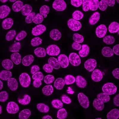


NucSpot® Live Cell Nuclear Stains are cell membrane-permeant DNA dyes that specifically stain nuclei in live or fixed cells. They have excellent specificity for DNA without the need for a wash step, with low toxicity for live cell imaging.
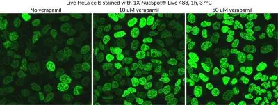
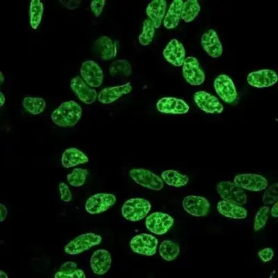
NucSpot® Live 488 stains nuclei with green fluorescence for the FITC channel, while NucSpot® Live 650 has far-red fluorescence for the Cy®5 channel. NucSpot® Live 650 dye also is compatible with super-resolution imaging by SIM or STED, and has been validated in STORM (Ref. 4). The dyes are supplied as 1000X stock solutions in DMSO, and are supplied with a vial of the efflux pump inhibitor verapamil for optional use, which may increase probe retention and live cell staining in some cell types. Note: NucSpot® Live 488 and NucSpot® Live 650 have dim blue fluorescence in the DAPI channel, and may not be suitable for multicolor imaging with blue probes.
NucSpot® Live Cell Nuclear Stains can be used to stain live gram-positive bacteria, but do not stain live gram-negative bacteria or yeast. See our Cellular Stains Table for more information on how our dyes stain various organisms.
Also see our wide selection of other Membrane and Organelle Stains, or download the Cellular Stains Brochure.
Cy Dye is a registered trademark of GE Healthcare.
| Dye | NucSpot® Live 488, NucSpot® Live 650 |
|---|---|
| Cellular localization | Nucleus |
| For live or fixed cells | For fixed cells, For live/intact cells |
| Assay type/options | Long term staining (24-72h), No-wash staining, Real-time imaging |
| Cell permeability | Membrane permeant |
| Colors | Green, Far-red |
| Fixation options | Fix after staining (formaldehyde), Fix after staining (methanol), Permeabilize after staining, Fix before staining (methanol), Fix before staining (formaldehyde) |
Contact our Customer Care, Sales & Scientific Assistance
Consult and asked questions about our products & services
Documentation of Technical & Safety Data Sheet, Guides and more...
We gladly support you by keeping you updated on our latest products and the developments around our services.
362 Upper Paya Lebar Rd, #07-15,
Singapore 534963
© 2015-2023 Atlantis Bioscience Pte Ltd. All rights reserved. Co Reg No: 201539516N