Product Description
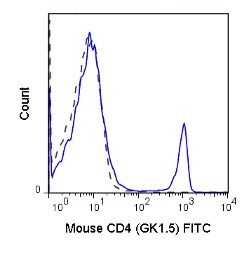
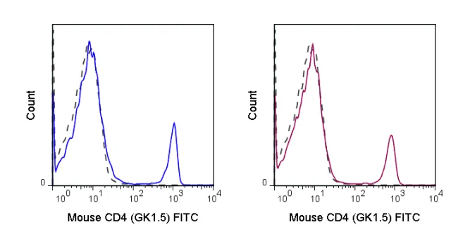
The GK1.5 antibody reacts with mouse CD4, a 55 kDa protein which acts as a co-receptor for the T cell receptor (TCR) in its interaction with MHC Class II molecules on antigen-presenting cells. The extracellular domain of CD4 binds to the beta-2 domain of MHC Class II, while its cytoplasmic tail provides a binding site for the tyrosine kinase lck, facilitating the signaling cascade that initiates T cell activation. CD4 is typically expressed on thymocytes, certain mature T cell populations such as Th17 and T regulatory (Treg) cells, as well as on dendritic cells.
The GK1.5 antibody is widely used as a phenotypic marker for CD4 expression. If used together, the GK1.5 antibody and an alternative antibody, Anti-Mouse CD4 clone RM4-5, will “compete” for binding, i.e. RM4-5 is able to block GK1.5 binding to cells. In contrast, the Anti-Mouse CD4 clone RM4-4 does not block binding of the GK1.5 antibody to cells (Arora S et al. 2006. Infect. Immun. 74: 4339-4348). The GK1.5 antibody is also reported to be cross-reactive with Syrian hamster CD4.
Recent Publications:
Higashino-Kameda M, Yabe-Wada T, Matsuba S, Takeda K, Anzawa K, Mochizuki T, Makimura K, Saijo S, Iwakura Y, Toga H and Nakamura A. 2015. Inflamm Res. DOI 10.1007/s00011-015-0910-1. (Flow Cytometry)
Product Details
Name | FITC Anti-Mouse CD4 (GK1.5) |
---|---|
Cat. No. | 35-0041 |
Alternative Names | L3T4, T4 |
Gene ID | 12504 |
Clone | GK1.5 |
Isotype | Rat IgG2b, κ |
Reactivity | Mouse |
Cross Reactivity | Syrian Hamster |
Format | FITC |
Application | Flow Cytometry |
Citations* | Taniguchi RT, DeVoss JJ, Moon JJ, Sidney J, Sette A, Jenkins MK, and Anderson MS. 2012. Proc. Natl. Acad. Sci. 109: 7847-7852. (in vivo depletion)
Wolkers MC, Gerlach, C, Arens R, Janssen EM, Fitzgerald P, Schumacher TN, Medema JP, Green DR, and Schoenberger SP. 2012. Blood. 119:798-804. (in vivo depletion) Lee L-F, Logronio K, Tu GH, Zhai W, Ni I, Mei L, Dilley J, Yu J, et al. 2012. Proc. Natl. Acad. Sci. 10.1073. (Flow cytometry). Thornton EE, Looney MR, Bose O, Sen D, Sheppard D, Locksley R, Huang X, and Krummel MF. 2012. J. Exp. Med. 10:1084. (Immunohistochemistry – OCT embedded frozen tissue) Stephen TL, Wilson BS, and Laufer TM. 2012. Proc. Natl. Acad. Sci. 109: 7415-7420. (Immunoprecipitation) Hammerbeck CD and Hooper JW. 2011. J. Virol. 85(19) 9929-9944. (Flow cytometry – Syrian hamster). Diamond MS, Kinder M, Matsushita H, Mashayekhi M, Dunn GP, Archambault JM, Lee H, Arthur CD, White JM, Kalinke U, Murphy KM, and Schreiber RD. 2011. J. Exp. Med. 208: 1989-2003. (in vitro blocking) Kao H, Lin J, Littman DR, Shaw AS, and Allen PM. 2008. J. Immunol. 181: 8248-8257. (Immunoprecipitation) Felix NJ, Donermeyer DL, Horvath S, Walters JJ, Gross M, suri A, and Allen PM. 2007. Nat. Immunol. 8:388-397. (in vitro blocking) |
Application Key:FC = Flow Cytometry; FA = Functional Assays; ELISA = Enzyme-Linked Immunosorbent Assay; ICC = Immunocytochemistry; IF = Immunofluorescence Microscopy; IHC = Immunohistochemistry; IHC-F = Immunohistochemistry, Frozen Tissue; IHC-P = Immunohistochemistry, Paraffin-Embedded Tissue; IP = Immunoprecipitation; WB = Western Blot; EM = Electron Microscopy
*Tonbo Biosciences tests all antibodies by flow cytometry. Citations are provided as a resource for additional applications that have not been validated by Tonbo Biosciences. Please choose the appropriate format for each application and consult the Materials and Methods section for additional details about the use of any product in these publications.