
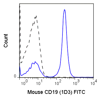
The 1D3 antibody reacts with mouse CD19, a 95 kDa glycoprotein which acts as a co-receptor, along with CD21 and CD81, in support of the functional B cell receptor (BCR). This complex provides antigen-specific recognition and subsequent activation of B cells to proliferate and differentiate into antibody-secreting cells (plasma cells) or memory B cells, which are crucial for secondary antigen encounter. CD19 is a lineage-differentiation marker, as its expression is detectable at the earliest B cell stages, through development, and is finally lost upon transition to mature plasma cells. The 1D3 antibody is widely used as a phenotypic marker for CD19 expression on B cells, as well as on dendritic cell subsets.
| Name | FITC Anti-Mouse CD19 (1D3) |
|---|---|
| Cat. No. | 35-0193 |
| Alternative Names | Leu-12, B4 |
| Gene ID | 12478 |
| Clone | 1D3 |
| Isotype | Rat IgG2a, κ |
| Reactivity | Mouse |
| Cross Reactivity | |
| Format | FITC |
| Application | Flow Cytometry |
| Citations* | Karki R, Sharma BR, Lee E, et al. Interferon regulatory factor 1 regulates PANoptosis to prevent colorectal cancer. JCI Insight. 2020;5(12):136720. Published 2020 Jun 18. doi:10.1172/jci.insight.136720.
Shiyang Li, John W. Bostick, Jian Ye, Joseph F. Urban, Jr., Dorina Avram, Liang Zhou. Aryl Hydrocarbon Receptor Signaling Cell Intrinsically Inhibits Intestinal Group 2 Innate Lymphoid Cell Function. Immunity. 2018 Nov 20;49(5):915-928.e5. doi: 10.1016/j.immuni.2018.09.015 Ghosn EEB, Yamamoto R, Hamanada S, Yang Y, Herzenberg LA, Nakauchi H, and Herzenberg LA. 2012. Proc. Natl. Acad. Sci. 109:5394-5398. (Flow cytometry) Raghavan S, Ostberg AK, Flach C-F, Ekman A, Blomquist M, Czerkinsky C, and Holmgren J. 2010. Infect. Immun. 78(10)4251-4260. (Immunohistochemistry – acetone fixed tissue) Togayachi A, Kozono Y, Ikehara Y, Ito H, et al. 2010. Proc. Natl. Acad. Sci. 107:11900-11905. (Immunoprecipitation) Poitrasson-Riviere M, Bienvenu B, Le Campion A, Becourt C, Martin B, and Lucas B. 2008. J. Immunol. 180:7294-7304. (Immunohistochemistry – frozen tissue) Lee Y, Haas KM, Gor DO, Ding X, Karp DR, Greenspan NS, Poe JC, and Tedder TF. 2005. J. Immunol. 175:8011-8023. (Immunoprecipitation) Bobbitt KR and Justement LB. 2000. J. Immunol. 165: 5588-5596. (in vitro stimulation, Immunoprecipitation) |
Application Key:FC = Flow Cytometry; FA = Functional Assays; ELISA = Enzyme-Linked Immunosorbent Assay; ICC = Immunocytochemistry; IF = Immunofluorescence Microscopy; IHC = Immunohistochemistry; IHC-F = Immunohistochemistry, Frozen Tissue; IHC-P = Immunohistochemistry, Paraffin-Embedded Tissue; IP = Immunoprecipitation; WB = Western Blot; EM = Electron Microscopy
*Tonbo Biosciences tests all antibodies by flow cytometry. Citations are provided as a resource for additional applications that have not been validated by Tonbo Biosciences. Please choose the appropriate format for each application and consult the Materials and Methods section for additional details about the use of any product in these publications.


Contact our Customer Care, Sales & Scientific Assistance
Consult and asked questions about our products & services
Documentation of Technical & Safety Data Sheet, Guides and more...
We gladly support you by keeping you updated on our latest products and the developments around our services.
362 Upper Paya Lebar Rd, #07-15,
Singapore 534963
© 2015-2023 Atlantis Bioscience Pte Ltd. All rights reserved. Co Reg No: 201539516N


