
| Cat No. | Size | Price |
|---|---|---|
| 30-5941-U025 | 25 µg | $30.00 |
| 30-5941-U100 | 100 µg | $84.00 |
| 30-5941-U100 | 500 µg | $140.00 |
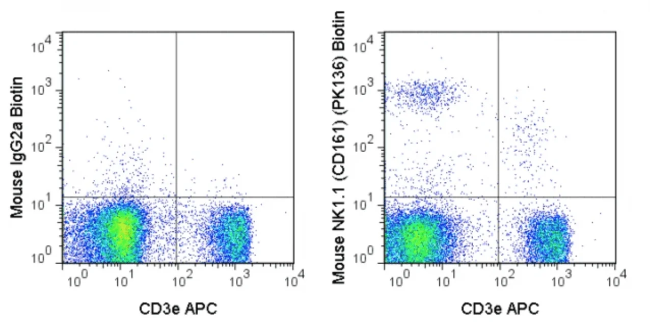
The PK136 antibody is specific for mouse NK1.1, a type II transmembrane lectin-like receptor and member of the killer cell lectin-like receptor (KLR) family. NK1.1 is prominently expressed on natural killer (NK) cells, and is correlated with NK cytotoxic effects toward virus-infected cells and tumor cells. NK1.1 is expressed on subsets of NKT cells in certain mouse strains (C57BL/6, FVB/N, and NZB), yet absent from others (AKR, BALB/c, CBA/J, C3H, DBA/1, DBA/2, NOD, SJL, and 129). Putative subsets of NK cells and their expression of NK1.1 antigen are of continuing interest, including NK1.1+/CD117+ (c-Kit) cells reported to be immunosuppressive for CD8+ T cells in a mechanism involving PD-1 and PD-L1 (Ehlers et al. 2012. Endocrinology. 10: 1247).
The PK136 antibody may be used for detection of NK1.1 expression on mouse strains including CE, B6, NZB, C58, Ma/My, ST, SJL, and FVB. The antibody is reported to react with an epitope common to NKR-P1B and NKR-P1C alloantigenic forms of NK1.1 (Carlyle et al. 2006. J. Immunol. 176: 7511-7524).
Product Details
| Name | Biotin Anti-Mouse NK1.1 (CD161) (PK136) |
|---|---|
| Cat. No. | 30-5941 |
| Alternative Names | CD161, NKR-P1C, Ly-55 |
| Gene ID | 17059 |
| Clone | PK136 |
| Isotype | Mouse IgG2a, κ |
| Reactivity | Mouse |
| Cross Reactivity | |
| Format | Biotin |
| Application | Flow Cytometry |
| Citations* | Krebs DL, Chehal MK, Sio A, Huntington ND, Da ML, Ziltener P, Inglese M, Kountouri N, Priatel JJ, Jones J, Tarlinton DM, Anderson GP, Hibbs ML, and Harder KW. 2012. J. Immunol. 188:5094-5105. (in vivo depletion)
Lubinski JM, Lazear HM, Awasthi S, Wang F, and Friedman HM. 2011. J. Virol. 85(7): 3239-3249. (in vivo depletion) Diamond MS, Kinder M, Matsushita H, Mashayekhi M, Dunn GP, Archambault JM, Lee H, Arthur CD, White JM, Kalinke U, Murphy KM, and Schreiber RD. 2011. J. Exp. Med. 208: 1989-2003. (in vivo depletion) Awasthi A, Samarakoon A, Chu H, Kamalakannan R, Quilliam LA, Chrzanowska-Wodnicka M, White GC, and Malarkannan S. 2010. J. Exp. Med. 207: 1923-1938. (in vitro activation) Coudert JD, Scarpellino L, Gros F, Vivier E, and Held W. 2008 Blood. 111: 3571-3578. (Immunoprecipitation) Ljutic B, Carlyle JR, Filipp D, Nakagawa R, Julius M, and Zuniga-Pflucker JC. 2005. J. Immunol. 174: 4789-4796. (Immunoprecipitation) Kanwar JR, Shen W-P, Kanwar RK, Berg RW, and Krissansen GW. 2001. J. Natl. Cancer Inst. 93: 1541-1552. (Immunohistochemistry – frozen tissue, Immunofluorescence microscopy – frozen tissue, in vivo depletion) |
Application Key:FC = Flow Cytometry; FA = Functional Assays; ELISA = Enzyme-Linked Immunosorbent Assay; ICC = Immunocytochemistry; IF = Immunofluorescence Microscopy; IHC = Immunohistochemistry; IHC-F = Immunohistochemistry, Frozen Tissue; IHC-P = Immunohistochemistry, Paraffin-Embedded Tissue; IP = Immunoprecipitation; WB = Western Blot; EM = Electron Microscopy
*Tonbo Biosciences tests all antibodies by flow cytometry. Citations are provided as a resource for additional applications that have not been validated by Tonbo Biosciences. Please choose the appropriate format for each application and consult the Materials and Methods section for additional details about the use of any product in these publications.
[accordions]
Contact our Customer Care, Sales & Scientific Assistance
Consult and asked questions about our products & services
Documentation of Technical & Safety Data Sheet, Guides and more...
We gladly support you by keeping you updated on our latest products and the developments around our services.
362 Upper Paya Lebar Rd, #07-15,
Singapore 534963
© 2015-2023 Atlantis Bioscience Pte Ltd. All rights reserved. Co Reg No: 201539516N


