Biotin Anti-Mouse IL-2 (JES6-5H4)

Biotin Anti-Mouse IL-2 (JES6-5H4)

Biotin Anti-Mouse IL-2 (JES6-5H4)

Add to cart

The JES6-5H4 antibody binds to mouse Interleukin-2 (IL-2), a 17 kDa cytokine that is secreted by activated T cells. This cytokine acts as an autocrine stimulatory factor for T cell proliferation, differentiation and survival, as well as being an effective activator of B cells and inducer of NK cell cytotoxic functions. IL-2 plays a key modulatory role in the differentiation of T cells toward either Th17 or T regulatory (Treg) cell types and is important for Treg cell function. The IL-2 receptor (IL-2R) is a complex consisting of an IL-2R alpha chain (CD25), along with the IL-2R beta chain (CD122) and the common gamma chain (CD132). IL-2 may also bind with low affinity to the T cell surface protein CD25 alone, although this interaction does not induce cell signaling.

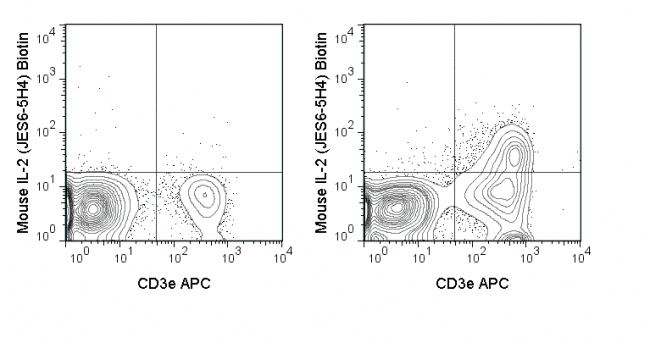
The JES6-5H4 antibody is extensively used for intracellular detection of IL-2 by flow cytometry, for detection of soluble cytokine in ELISA, and as a neutralizing reagent in vitro and in vivo.

Product Details

Name Biotin Anti-Mouse IL-2 (JES6-5H4)
Cat. No. 30-7021
Alternative Names Interleukin-2
Gene ID 16183
Clone JES6-5H4
Isotype Rat IgG2b, κ
Reactivity Mouse
Cross Reactivity
Format Biotin
Application Flow Cytometry
Citations* Attridge K, Wang CJ, Wardzinski L, Kenefeck R, Chamberlain JL, Manzotti C, Kopf M, and Walker LSK. 2012. Blood. 119: 4656-4664. (Flow cytometry)

Helbig C, Gentek R, Backer RA, de Souza Y, Derks IAM, Eldering E, Wagner K, Jankovic D, Gridley T, Moerland PD, Flavell RA, and Amsen D. 2012. Proc. Natl. Acad. Sci. 109: 9041-9046. (ELISA – detection)

Chen Q, Kiim YC, Laurence A, Punkosdy GA, and Shevach EM. 2011. J. Immunol. 186: 6329-6337. (in vivo blocking)

Weber KS, Hildner K, Murphy KM and Allen PM. 2010 J. Immunol. 185: 2836-2846. (ELISA)

Wang C, Morley SC, Donermeyer D, Peng I, Lee WP, Devoss J, Damilenko DM, Lin Z, Zhang J, Zhou J, Allen PM, and Brown EJ. 2010. J. Immunol. 185: 7487-7497. (Immunocytochemistry/Immunofluorescence microscopy)

Cardona AE and Teale JM. 2002. J. Immunol. 169: 3163-3171. (Immunohistochemistry – frozen tissue)

Woo AL, Gildea LA, Tack LM, Miller ML, Spicer A, Millhorn DE, Finkelman FD, Dassett DJ, and Shull GE. 2002. J. Biol. Chem. 277: 49036-49046. (in vivo blocking)

Application Key:FC = Flow Cytometry; FA = Functional Assays; ELISA = Enzyme-Linked Immunosorbent Assay; ICC = Immunocytochemistry; IF = Immunofluorescence Microscopy; IHC = Immunohistochemistry; IHC-F = Immunohistochemistry, Frozen Tissue; IHC-P = Immunohistochemistry, Paraffin-Embedded Tissue; IP = Immunoprecipitation; WB = Western Blot; EM = Electron Microscopy

*Tonbo Biosciences tests all antibodies by flow cytometry. Citations are provided as a resource for additional applications that have not been validated by Tonbo Biosciences. Please choose the appropriate format for each application and consult the Materials and Methods section for additional details about the use of any product in these publications.

Technical Data Sheet                   View All Formats                      Request Custom/Bulk