
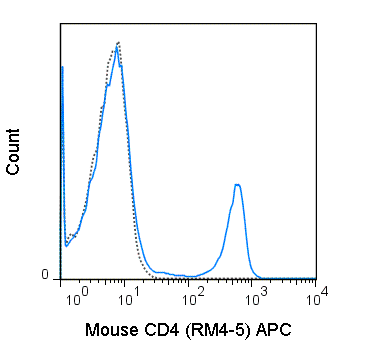
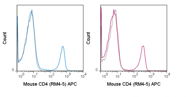
The RM4-5 antibody reacts with mouse CD4, a 55 kDa protein which acts as a co-receptor for the T cell receptor (TCR) in its interaction with MHC Class II molecules on antigen-presenting cells. The extracellular domain of CD4 binds to the beta-2 domain of MHC Class II, while its cytoplasmic tail provides a binding site for the tyrosine kinase lck, facilitating the signaling cascade that initiates T cell activation. CD4 is typically expressed on thymocytes, certain mature T cell populations such as Th17 and T regulatory (Treg) cells, as well as on dendritic cells.
The RM4-5 antibody is widely used as a phenotypic marker for CD4 expression. If used together, the RM4-5 antibody and an alternative antibody, Anti-Mouse CD4 clone GK1.5, will “compete” for binding, i.e. RM4-5 antibody is able to block GK1.5 antibody binding to cells. In contrast, RM4-5 antibody does not block the binding of Anti-Mouse CD4 clone RM4-4 to cells.
| Name | APC Anti-Mouse CD4 (RM4-5) |
|---|---|
| Cat. No. | 20-0042 |
| Alternative Names | L3T4, T4 |
| Gene ID | 12504 |
| Clone | RM4-5 |
| Isotype | Rat IgG2a, κ |
| Reactivity | Mouse |
| Cross Reactivity | |
| Format | APC |
| Application | Flow Cytometry |
| Citations* | Willinger T and Flavell RA. 2012. Proc. Natl. Acad. Sci. 109:8670-8675. (Flow cytometry)
Stephen TL, Wilson BS, and Laufer TM. 2012. Proc. Natl. Acad. Sci. 109: 7415-7420. (Immunofluorescence microscopy) Poitrasson-Riviere M, Bienvenu B, Le Campion A, Becourt C, Martin B, and Lucas B. 2008. J. Immunol. 180:7294-7304. (Immunohistochemistry – paraffin embedded tissue) Sorg H, Lorch B, Jaster R, Fitzner B, Ibrahim S, Holzhueter S, Nizze H, and Vollmar B. 2008. Am. J. Physio. Gastrointest. Liver Physiol. 295: G1274-1280. (Immunohistochemistry – paraffin embedded tissue) Menke J, Lucas JA, Zeller GC, Keir ME, Huang XR, Tsuboi N, Mayadas TN, Lan HY, Sharpe AH, and Kelley VR. 2007. J. Immunol. 179: 7466-7477. (Immunohistochemistry – frozen tissue) Irie J, Wu Y, Wicker LS, Rainbow D, Nalesnik MA, Hirsch R, Peterson LB, Leung PS, Cheng C, Mackay IR, Gershwin ME, and Ridgway WM. 2006. J Exp Med. 203(5):1209-19. (Immunohistochemistry – frozen tissue) Bosselut R, Zhang W, Ashe JM, Kopacz JL, Samelson LE, and Singer A. 1999. J. Exp. Med. 190: 1517-1526. (Immunoprecipitation) Shi Y, Kaliyaperumal A, Lu L, Southwood S, Sette A, Michaels MA, and Datta SK. 1998. J. Exp. Med. 187:367-378. (Blocking) Whiteland JL, Nicholls SM, Shimeld C, Easty DL, Williams NA, and Hill TJ. 1995. J. Histochem. Cytochem. 43:313-320. (Immunohistochemistry – frozen tissue, zinc-fixed paraffin embedded tissue) |
Application Key:FC = Flow Cytometry; FA = Functional Assays; ELISA = Enzyme-Linked Immunosorbent Assay; ICC = Immunocytochemistry; IF = Immunofluorescence Microscopy; IHC = Immunohistochemistry; IHC-F = Immunohistochemistry, Frozen Tissue; IHC-P = Immunohistochemistry, Paraffin-Embedded Tissue; IP = Immunoprecipitation; WB = Western Blot; EM = Electron Microscopy
*Tonbo Biosciences tests all antibodies by flow cytometry. Citations are provided as a resource for additional applications that have not been validated by Tonbo Biosciences. Please choose the appropriate format for each application and consult the Materials and Methods section for additional details about the use of any product in these publications.


Contact our Customer Care, Sales & Scientific Assistance
Consult and asked questions about our products & services
Documentation of Technical & Safety Data Sheet, Guides and more...
We gladly support you by keeping you updated on our latest products and the developments around our services.
362 Upper Paya Lebar Rd, #07-15,
Singapore 534963
© 2015-2023 Atlantis Bioscience Pte Ltd. All rights reserved. Co Reg No: 201539516N


