
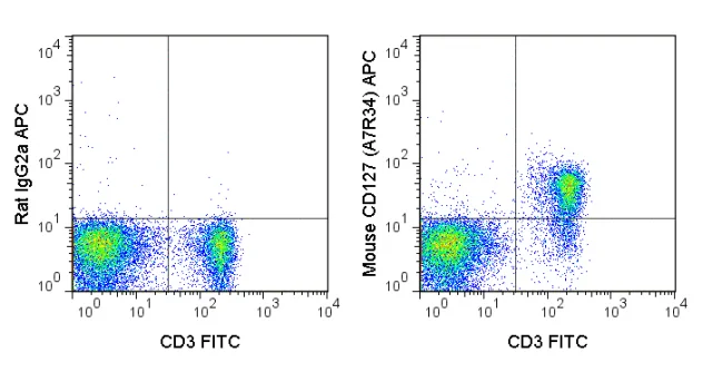
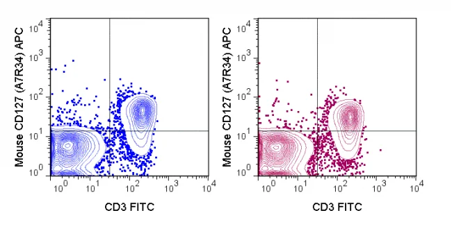
The A7R34 antibody is specific for mouse CD127, a 60-90 kDa cell surface protein also known as the Interleukin-7 Receptor alpha chain, or IL-7R alpha. CD127 is typically expressed at the cell surface as a heterodimer with the common gamma chain (CD132). This complex acts as the functional receptor for IL-7, a cytokine important in T and B cell development, and in mature T cell homeostasis. A second cytokine known as Thymic Stromal Lymphopoietin (TSLP) also binds to a receptor complex of CD127 and the TSLPR chain to trigger activation of dendritic cells, and is involved in B cell development, allergy and autoimmunity.
The A7R34 antibody may be used as a phenotypic marker for CD127 on immature B cells, on subsets of thymocytes which are double negative (CD4-CD8- ) or single positive (CD4+ or CD8+), and at low levels on mature, peripheral T cells. CD127 is a key marker, when used in combination with CD4 and CD25, to distinguish Treg and effector/memory Treg populations known as T(REM).
| Name | APC Anti-Mouse CD127 (IL-7Ra) (A7R34) |
|---|---|
| Cat. No. | 20-1271 |
| Alternative Names | Interleukin-7 Receptor alpha, IL-7Ra |
| Gene ID | 16172 |
| Clone | A7R34 |
| Isotype | Rat IgG2a, κ |
| Reactivity | Mouse |
| Cross Reactivity | |
| Format | APC |
| Application | Flow Cytometry |
| Citations* | Shiyang Li, John W. Bostick, Jian Ye, Joseph F. Urban, Jr., Dorina Avram, Liang Zhou. Aryl Hydrocarbon Receptor Signaling Cell Intrinsically Inhibits Intestinal Group 2 Innate Lymphoid Cell Function. Immunity. 2018 Nov 20;49(5):915-928.e5. doi: 10.1016/j.immuni.2018.09.015
Thaventhiran JED, Hoffmann A, Magiera L, de la Roche M, Lingel H, Brunner-Weinzierl M, and Fearon DT. 2012. Proc. Natl. Acad. Sci. 10.1073. (Flow cytometry). Jin J, Goldschneider I, and Lai L. 2011. J. Immunol. 186: 1915-1922. (in vivo activation) Vondenhoff MF, Greuter M, Goverse G, Elewaut D, Dewint P, Ware CF, Hoorweg K, Kraal G, and Mebius RE. 2009. J. Immunol. 182(9): 5439-5445. (Immunofluorescence microscopy – frozen tissue) Leithauser F, Meinhardt-Krajina T, Fink K, Wotschke B, Moller P and Reimann J. 2006. Am. J. Pathol. 168(6): 1898-1909. (Immunohistochemistry – frozen tissue) Seddon B and Zamoyska R. 2002. J. Immunol. 169: 2997-3005. (in vivo activation) Sudo T, Nishikawa S, Ohno N, Akiyama N, Tamakoshi M, Yoshida H and Nishikawa S-I. 1993. Proc. Natl. Acad. Sci. 90: 9125-9129. (in vitro and in vivo blocking; Immunoprecipitation) |
Application Key:FC = Flow Cytometry; FA = Functional Assays; ELISA = Enzyme-Linked Immunosorbent Assay; ICC = Immunocytochemistry; IF = Immunofluorescence Microscopy; IHC = Immunohistochemistry; IHC-F = Immunohistochemistry, Frozen Tissue; IHC-P = Immunohistochemistry, Paraffin-Embedded Tissue; IP = Immunoprecipitation; WB = Western Blot; EM = Electron Microscopy
*Tonbo Biosciences tests all antibodies by flow cytometry. Citations are provided as a resource for additional applications that have not been validated by Tonbo Biosciences. Please choose the appropriate format for each application and consult the Materials and Methods section for additional details about the use of any product in these publications.


Contact our Customer Care, Sales & Scientific Assistance
Consult and asked questions about our products & services
Documentation of Technical & Safety Data Sheet, Guides and more...
We gladly support you by keeping you updated on our latest products and the developments around our services.
362 Upper Paya Lebar Rd, #07-15,
Singapore 534963
© 2015-2023 Atlantis Bioscience Pte Ltd. All rights reserved. Co Reg No: 201539516N


