
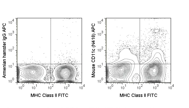
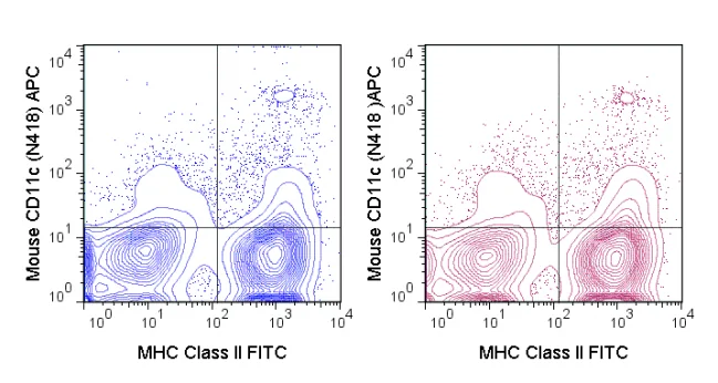
The N418 antibody reacts with mouse CD11c, also known as integrin αalpha X. This 150 kDa cell surface glycoprotein is part of a family of integrin αreceptors that mediate adhesion between ≥ ≥ ≥ cells (cell-cell) and components of the extracellular matrix, e.g. fibrinogen (cell-matrix). In addition, integrin αs are active signaling receptors which recruit leukocytes to inflammatory sites and promote cell activation. Complete, functional integrin αreceptors consist of distinct combinations of integrin αchains which are differentially expressed. integrin αalpha X (CD11c) assembles with integrin αbeta-2 (CD18) into a receptor complex known as CR4 which can bind and induce signaling through ICAMs and VCAM-1 on endothelial cells and can also facilitate removal of iC3b bearing foreign cells.
The N418 antibody is widely used as a marker for CD11c expression on dendritic cells (DC), often in parallel with markers for CD11b, for identification of developmental stages and mature subsets of this cell type. CD11c is prominently expressed on tissue macrophages, and is also detected on some types of activated T cells and intestinal intraepithelial lymphocytes (IEL).
Recent Publications:
Tani Y, Isobe Y, Imoto Y, Segi-Nishida E, Sugimoto Y, Arai H, & Arita M. 2014. FASEB J. fj.14-251132. (Flow Cytometry)
| Name | APC Anti-Mouse CD11c (N418) |
|---|---|
| Cat. No. | 20-0114 |
| Alternative Names | p150, integrin α?X, Itgax, CR4 |
| Gene ID | 16411 |
| Clone | N418 |
| Isotype | Armenian Hamster IgG |
| Reactivity | Mouse |
| Cross Reactivity | |
| Format | APC |
| Application | Flow Cytometry |
| Citations* | Guerriero JL, Ditsworth D, Catanzaro JM, Sabino G, Furie MB, Kew RR, Crawford HC, and Zong W-X. 2011. J. Immunol. 186: 3517-3526. (Immunohistochemistry – paraffin embedded tissue)
Grewal JS, Pilgrim MJ, Grewal S, Kasman L, Werner P, Bruorton ME, London SD, and London L. 2011. FASEB J. 25:1680-1696. (Immunofluorescence microscopy – frozen tissue) Sadhu C, Ting HJ, Lipsky B, Hensley K, Garcia-Martinez LF, Simon SI, and Staunton DE. 2007. J. Leukoc. Biol. 81: 1395-1403. (in vitro blocking) Hagnerud, S, Manna PP, Cella M, Stenberg A, Frazier WA, Colonna M, and Oldenborg P-A. 2006. J. Immunol. 5772-5778. (Immunofluorescence microscopy – frozen tissue) Finkelman FD, Lees A, Birnbaum R, Gause WC, and Morris SC. 1996. J. Immunol. 157: 1406-1414. (in vivo activation) Huleatt JW and Lefrancois L. 1995. J. Immunol. 154: 5684-5693. (Immunoprecipitation) Metlay JP, Witmer-Pack MD, Agger R, Crowley MT, Lawless D, and Steinman RM. 1990. J. Exp. Med. 171: 1753. (Immunoprecipitation) Specific References: |
Application Key:FC = Flow Cytometry; FA = Functional Assays; ELISA = Enzyme-Linked Immunosorbent Assay; ICC = Immunocytochemistry; IF = Immunofluorescence Microscopy; IHC = Immunohistochemistry; IHC-F = Immunohistochemistry, Frozen Tissue; IHC-P = Immunohistochemistry, Paraffin-Embedded Tissue; IP = Immunoprecipitation; WB = Western Blot; EM = Electron Microscopy
*Tonbo Biosciences tests all antibodies by flow cytometry. Citations are provided as a resource for additional applications that have not been validated by Tonbo Biosciences. Please choose the appropriate format for each application and consult the Materials and Methods section for additional details about the use of any product in these publications.


Contact our Customer Care, Sales & Scientific Assistance
Consult and asked questions about our products & services
Documentation of Technical & Safety Data Sheet, Guides and more...
We gladly support you by keeping you updated on our latest products and the developments around our services.
362 Upper Paya Lebar Rd, #07-15,
Singapore 534963
© 2015-2023 Atlantis Bioscience Pte Ltd. All rights reserved. Co Reg No: 201539516N


