
| Cat No. | Size | Price |
|---|---|---|
| 20-0087-T025 | 25 tests | $110.00 |
| 20-0087-T100 | 100 tests | $285.00 |
| 20-0087-T100 | 500 tests | $1282.00 |
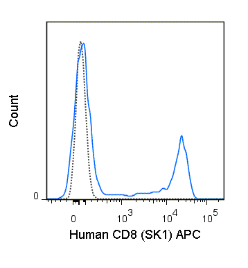
The SK1 antibody is specific for the 32-34 kDa alpha chain of human CD8, known as CD8a or CD8 alpha. CD8a can form a homodimer (CD8 alpha-alpha), but is more commonly expressed as a heterodimer with a second chain known as CD8b or CD8 beta. CD8 acts as a co-receptor for antigen recognition and subsequent T cell activation that is initiated upon binding of the T cell receptor (TCR) to antigen-bearing MHC Class I molecules. The cytoplasmic domains of CD8 provide binding sites for the tyrosine kinase lck, facilitating intracellular signaling events that lead to T cell activation, development, and cytotoxic effector functions. CD8+ cytotoxic T cells (CTLs) play an important role in inducing cell death of tumor cells, as well as cells infected by virus, bacteria or parasites.
The SK1 antibody is widely used as a phenotypic marker for CD8 on cytotoxic T cells, thymocytes, as well as on certain cell types that do not also express the TCR, including some NK cells and lymphoid dendritic cells. It is cross-reactive with CD8 in several non-human species, including Baboon, Chimpanzee, Cynomolgus and Rhesus. If used together with an alternative Anti-Human CD8a clone, RPA-T8, the SK1 antibody will not block binding of RPA-T8 to CD8a.
Product Details
| Name | APC Anti-Human CD8 (SK1) |
|---|---|
| Cat. No. | 20-0087 |
| Alternative Names | CD8 alpha, leu-2a |
| Gene ID | 925 |
| Clone | SK1 |
| Isotype | Mouse IgG1, kappa |
| Reactivity | Human |
| Cross Reactivity | African Green, Baboon, Chimpanzee, Cynomolgus, Rhesus, Pigtailed Macaque, Sooty Mangabey |
| Format | APC |
| Application | Flow Cytometry |
| Citations* | Campanelli R, Palermo B, Garbelli S, Mantovani S, Lucchi P, Necker A, Lantelme E and Giachino C. 2002. Int Immunol. 14(1): 39-44. (Flow Cytometry)
Walter S, Herrgen L, Schoor O, Jung G, Wernet D, Buhring H-J, Rammensee H-G and Stevanovic S. 2003. J. Immunol. 171(10): 4974-4978. (Flow Cytometry) Cervasi B, Paiardinin M, Serafini S, Fraternale A, Menotta M, Engram J, Lawson B, Staprans I, Piedimonte G, Perno CF, Silvestri G and Magnani M. 2006. J. Virol. 80(21): 10335-10345. (Flow Cytometry – Sooty Mangabeys) Verstrepen BE, Verschoor EJ, Fagrouch ZC, Mooij P, de Groot NG, Bontrop RE, Bogers WM, Heeney JL and Koopman G. 2014. PLoS ONE 9(4): e95103. doi:10.1371/journal.pone.0095103. (Flow Cytometry – Chimpanzee) Permar SR, Klumpp SA, Mansfield KG, Kim W-K, Gorgone DA, Lifton MA, Williams KC, Schmitz JE, Reimann KA, Axthelm MK, Polack FP, Griffin DE and Letvin NL. 2003. J. Virol. 77(7): 4396-4400. (Flow Cytometry – Rhesus) Ahmed AFK, Ohtani H, Nio M, Fuanki N, Shimaoka S, Nagura H and Ohi R. 2001. J. Pathol. 193(3): 383-389. (Immunohistochemistry) Bukowska-Straková K, Baran J, Gawlicka M and Kowalczyk, D. 2006. Folia Histochemica et Cytobiologica 44(3): 179-183. |
Application Key:FC = Flow Cytometry; FA = Functional Assays; ELISA = Enzyme-Linked Immunosorbent Assay; ICC = Immunocytochemistry; IF = Immunofluorescence Microscopy; IHC = Immunohistochemistry; IHC-F = Immunohistochemistry, Frozen Tissue; IHC-P = Immunohistochemistry, Paraffin-Embedded Tissue; IP = Immunoprecipitation; WB = Western Blot; EM = Electron Microscopy
*Tonbo Biosciences tests all antibodies by flow cytometry. Citations are provided as a resource for additional applications that have not been validated by Tonbo Biosciences. Please choose the appropriate format for each application and consult the Materials and Methods section for additional details about the use of any product in these publications.
[accordions]
Contact our Customer Care, Sales & Scientific Assistance
Consult and asked questions about our products & services
Documentation of Technical & Safety Data Sheet, Guides and more...
We gladly support you by keeping you updated on our latest products and the developments around our services.
362 Upper Paya Lebar Rd, #07-15,
Singapore 534963
© 2015-2023 Atlantis Bioscience Pte Ltd. All rights reserved. Co Reg No: 201539516N


