
The OKT4 antibody reacts with human CD4, a 59 kDa protein which acts as a co-receptor for the T cell receptor (TCR) in its interaction with MHC Class II molecules on antigen-presenting cells. The extracellular domain of CD4 binds to the beta-2 domain of MHC Class II, while its cytoplasmic tail provides a binding site for the tyrosine kinase lck, facilitating the signaling cascade that initiates T cell activation. CD4, and co-receptors CCR5 and CXCR4, may also be utilized by HIV-1 to enter T cells. Human CD4 is typically expressed on thymocytes, some mature T cell populations such as Th17 and T regulatory (Treg) cells, as well as on dendritic cells.
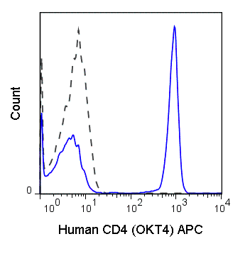
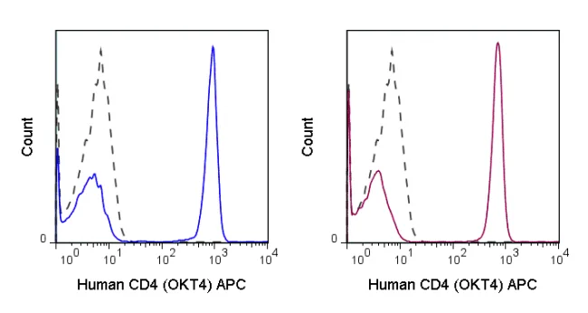
The OKT4 antibody is widely used as a phenotypic marker for CD4 expression. It is cross-reactive with CD4 in several non-human species, including Chimpanzee, Cynomolgus and Rhesus. This antibody recognizes a different epitope, and thus does not block binding of, the alternative Anti-Human CD4 antibody clone RPA-T4 (Reinherz EL, et al. 1979. Proc. Natl. Acad. Sci. 76:4061-4065)
| Name | APC Anti-Human CD4 (OKT4) |
|---|---|
| Cat. No. | 20-0048 |
| Alternative Names | Leu-3, T4 |
| Gene ID | 920 |
| Clone | OKT4 |
| Isotype | Mouse IgG2b, kappa |
| Reactivity | Human |
| Cross Reactivity | Chimpanzee, Cynomolgus, Rhesus |
| Format | APC |
| Application | Flow Cytometry |
| Citations* | Chen CY, Huang D, Yao S, Halliday L, Zeng G, Wang RC and Chen ZW. 2012. J. Immunol. 188:4278-4288. (in vivo depletion – macaque)
Bagnara D, Kaufman MS, Calissano C, et al. 2011. Blood. 117: 5463-5472. (in vivo depletion) Ciczora Y, Callens N, Seron K, Rouille Y, and Dubuisson J. 2010. J. Gen. Virol. 91:404-414. (Immunofluorescence microscopy; Western Blot) Nguyen V, Cao L, Lin JT, Hung N, Ritz A, Yu K, Jianu R, Ulin SP, Raphael BJ, Laidlaw DH, Brossay L, and Salomon AR. 2009. Mol. Cell. Proteomics. 8: 2418-2431. (in vitro activation) daSilva LLP, Sougrat R, Burgos PV, Janvier K, Mattera R, and Bonifacino JS. 2009. J. Virol. 83: 6578-6590 (Immunoprecipitation) Balla-Jhagjhoorsingh S, Koopman G, Mooij P, Haaksma TGM, Teeuwsen VJP, Bontrop RE, and Heeney JL. 1999. J. Immunol. 162: 2308-2314. (Immunocytochemistry /Immunofluorescence microscopy – Chimpanzee) Bour S, Boulerice F, and Wainberg MA. 1991. J. Virol. 65(12) : 6387-6396. (Immunoprecipitation)Watanabe M, Ringler DJ, Fultz PN, MacKey JJ, Boyson JE, Levine CG, and Letvin NL. 1991. J. Virol. 65: 3344-3348. (Flow cytometry – Chimpanzee) |
Application Key:FC = Flow Cytometry; FA = Functional Assays; ELISA = Enzyme-Linked Immunosorbent Assay; ICC = Immunocytochemistry; IF = Immunofluorescence Microscopy; IHC = Immunohistochemistry; IHC-F = Immunohistochemistry, Frozen Tissue; IHC-P = Immunohistochemistry, Paraffin-Embedded Tissue; IP = Immunoprecipitation; WB = Western Blot; EM = Electron Microscopy
*Tonbo Biosciences tests all antibodies by flow cytometry. Citations are provided as a resource for additional applications that have not been validated by Tonbo Biosciences. Please choose the appropriate format for each application and consult the Materials and Methods section for additional details about the use of any product in these publications.


Contact our Customer Care, Sales & Scientific Assistance
Consult and asked questions about our products & services
Documentation of Technical & Safety Data Sheet, Guides and more...
We gladly support you by keeping you updated on our latest products and the developments around our services.
362 Upper Paya Lebar Rd, #07-15,
Singapore 534963
© 2015-2023 Atlantis Bioscience Pte Ltd. All rights reserved. Co Reg No: 201539516N


