
| Cat No. | Size | Price |
|---|---|---|
| 20-0039-T025 | 25 tests | $41.00 |
| 20-0039-T100 | 100 tests | $84.00 |
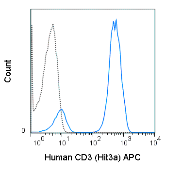
The Hit3a antibody is specific for human CD3e, also known as CD3 epsilon, a 20 kDa subunit of the T cell receptor complex, along with CD3 gamma and CD3 delta. These integral membrane protein chains assemble with additional chains of the T cell receptor (TCR), as well as CD3 zeta chain, to form the T cell receptor – CD3 complex. Together with co-receptors CD4 or CD8, the complex serves to recognize antigens bound to MHC molecules on antigen-presenting cells. These interactions promote T cell receptor signaling (T cell activation), inducing cell proliferation, differentiation, production of cytokines or activation-induced cell death. CD3 is differentially expressed during thymocyte-to-T cell development and on all mature T cells.
The Hit3a antibody is a widely used phenotypic marker for human T cells. In addition, binding/cross-linking of Hit3a antibody to CD3e can induce cell activation. The antibody has also been demonstrated to be cross-reactive with Chimpanzee CD3. Please choose the appropriate format for each application.
| Name | APC Anti-Human CD3 (Hit3a) |
|---|---|
| Cat. No. | 20-0039 |
| Alternative Names | Leu-4, T3 |
| Gene ID | 916 |
| Clone | Hit3a |
| Isotype | Mouse IgG2a, κ |
| Reactivity | Human |
| Cross Reactivity | Chimpanzee |
| Format | APC |
| Application | Flow Cytometry |
| Citations* | Lesourne R, Zvezdova E, Song K-D, El-Khoury D, Uehara S, Barr VA, Samelson LE and Love PE. 2012. J. Immunol. 189: 1154-1161. (in vitro activation)
Knyazhitsky M, Moas E, Shaginov E, Luria A, and Braiman A. 2012. J. Biol. Chem. 287: 19725-19735. (in vitro activation) Ge Shuwang, Hertel B, Emden SH, Beneke J, Menne J, Haller H, and von Vietinghoff S. 2012. Nephrol. Dial. Transplant. 27: 2768-2772. (Immunofluorescence microscopy) Soto PC, Stein LL, Hurtado-Ziola N, Hedrick SM, and Varki A. 2010. J. Immunol. 184: 4185-4195. (Flow cytometry – Chimpanzee) Westermann J, Bode U, Sahle A, Speck U, Karin N, Bell EB, Kalies K, and Gebert A. 2005. J. Immunol. 174: 2517-2524. (Immunohistochemistry – frozen tissue) Mukouyama H, Janzen NK, Hernandez JM, Lam JS, Caliliw R, Wang AY, Figlin RA, Belldegrun AS, and Zeng G. 2004. Clin. Cancer Res. 10: 1421-1429. (in vitro blocking) |
Application Key:FC = Flow Cytometry; FA = Functional Assays; ELISA = Enzyme-Linked Immunosorbent Assay; ICC = Immunocytochemistry; IF = Immunofluorescence Microscopy; IHC = Immunohistochemistry; IHC-F = Immunohistochemistry, Frozen Tissue; IHC-P = Immunohistochemistry, Paraffin-Embedded Tissue; IP = Immunoprecipitation; WB = Western Blot; EM = Electron Microscopy
*Tonbo Biosciences tests all antibodies by flow cytometry. Citations are provided as a resource for additional applications that have not been validated by Tonbo Biosciences. Please choose the appropriate format for each application and consult the Materials and Methods section for additional details about the use of any product in these publications.
[accordions]
Contact our Customer Care, Sales & Scientific Assistance
Consult and asked questions about our products & services
Documentation of Technical & Safety Data Sheet, Guides and more...
We gladly support you by keeping you updated on our latest products and the developments around our services.
362 Upper Paya Lebar Rd, #07-15,
Singapore 534963
© 2015-2023 Atlantis Bioscience Pte Ltd. All rights reserved. Co Reg No: 201539516N


