
| Cat No. | Size | Price |
|---|---|---|
| 20-0118-T025 | 25 tests | $38.00 |
| 20-0118-T100 | 100 tests | $115.00 |
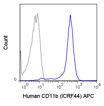
The ICRF44 antibody reacts with human CD11b, also known as integrin αalpha M. This 165-170 kDa cell surface glycoprotein is part of a family of integrin αreceptors that mediate adhesion between ≥ ≥ ≥ cells (cell-cell) and components of the extracellular matrix, e.g. fibrinogen (cell-matrix). In addition, integrin αs are active signaling receptors which recruit leukocytes to inflammatory sites and promote cell activation. Complete, functional integrin αreceptors consist of distinct combinations of integrin αchains which are differentially expressed. integrin αalpha M (CD11b) assembles with integrin αbeta-2 (CD18) into a receptor known as Macrophage Antigen-1 (Mac-1) or complement receptor type 3 (CR3). This receptor binds and induces intracellular signaling through ICAM-1, ICAM-2, ICAM-3 and ICAM-4 on endothelial cells and can also facilitate removal of iC3b bearing foreign cells.
The ICRF44 antibody is widely used as a marker for CD11b expression on macrophages, granulocytes, and subsets of NK cells. It is reported to be cross-reactive with a number of non-human species including Baboon, Chimpanzee, Cynomolgus, Rhesus and Swine.
| Name | APC Anti-Human CD11b (ICRF44) |
|---|---|
| Cat. No. | 20-0118 |
| Alternative Names | Mac-1, integrin ααM, Itgam, CR3 |
| Gene ID | 3684 |
| Clone | ICRF44 |
| Isotype | Mouse IgG1, kappa |
| Reactivity | Human |
| Cross Reactivity | Baboon, Chimpanzee, Cynomolgus, Rhesus, Swine |
| Format | APC |
| Application | Flow Cytometry |
| Citations* | Feng C, Zhang L, Almulki L, Faez S, Whitford M, Hafezi-Moghadam A, and Cross AS. 2011. J. Leukoc. Biol. 90:313-321. (Immunoprecipitation)
Chang WLW and Barry PA. 2010. Proc. Natl. Acad. Sci. 107:22647-2652. (Flow cytometry – Rhesus macaque) Jerke U, Rolle S, Dittmar G, Bayat B, Santoso S, Sporbert A, Luft F, and Kettritz R. 2010. J. Biol. Chem. 286:7070-7081. (in vitro blocking) Moreau A, Hill M, Thebault P, Deschamps JY, Chiffoleau E, Chauveau C, Moullier P, Anegon I, Alliot-Licht B, and Cuturi MC. 2009. FASEB J. 23:3070-3077. (Flow cytometry – cynomolgus macaque) Sengoku K, Takuma N, Miyamoto T, Horikawa M, and Ishikawa M. 2004. Hum. Reprod. 19: 639-644. (Immunofluorescence microscopy) David A, Kacher Y, Specks U, and Aviram I. 2003. J. Leukoc. Biol. 74:551-557. (Western blot)Rezzonico R, Imbert V, Chicheportiche R, and Dayer J-M. 2001. Blood. 97: 2932-2940. (in vitro activation) |
Application Key:FC = Flow Cytometry; FA = Functional Assays; ELISA = Enzyme-Linked Immunosorbent Assay; ICC = Immunocytochemistry; IF = Immunofluorescence Microscopy; IHC = Immunohistochemistry; IHC-F = Immunohistochemistry, Frozen Tissue; IHC-P = Immunohistochemistry, Paraffin-Embedded Tissue; IP = Immunoprecipitation; WB = Western Blot; EM = Electron Microscopy
*Tonbo Biosciences tests all antibodies by flow cytometry. Citations are provided as a resource for additional applications that have not been validated by Tonbo Biosciences. Please choose the appropriate format for each application and consult the Materials and Methods section for additional details about the use of any product in these publications.
[accordions]
[accordion title=”Protocols”]Technical Date Sheet[/accordion]
[accordions]
[accordion title=”SDS”] sds-70050[/accordion]
[accordions]
[accordion title=”Supporting Documents”]Cellular Stains,
Nerve Terminal Probes[/accordion]
Contact our Customer Care, Sales & Scientific Assistance
Consult and asked questions about our products & services
Documentation of Technical & Safety Data Sheet, Guides and more...
We gladly support you by keeping you updated on our latest products and the developments around our services.
362 Upper Paya Lebar Rd, #07-15,
Singapore 534963
© 2015-2023 Atlantis Bioscience Pte Ltd. All rights reserved. Co Reg No: 201539516N


