[vc_row][vc_column][vc_row_inner][vc_column_inner width=”1/6″][/vc_column_inner][vc_column_inner width=”2/3″][vc_column_text]
Cellular Imaging
[/vc_column_text][/vc_column_inner][vc_column_inner width=”1/6″][/vc_column_inner][/vc_row_inner][vc_separator][/vc_column][/vc_row][vc_row][vc_column][vc_column_text]
Cell biology was discovered through the endeavour of biologists by using only a brightfield microscope in the 1600s. This hardship of biologists’ work and unresolved questions has inspired scientists to develop new cellular imaging techniques such as phase-contrast microscopy, confocal microscopy, fluorescence microscopy, live-cell microscopy, and super resolution microscopy.
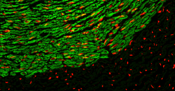
Phase-contrast microscopy allows scientists to visualize living cells in their natural condition through high contrast and high-resolution images. Confocal microscopy has the ability to control depth-of-field, collect serial optical sections from thick specimens, reduce background noise, and create 3D images from internal structures of the cells. The fluorescence microcopy allows scientists to label and identify the structure of interest in fixed and living cells by using specific antibodies/dyes and/or cellular stains. To obtain real-time biological processes, time-lapse live cell imaging was developed for studying the dynamic of internal structures and cellular processes. The observation of the dynamic changes provides more insights to the processes of the cell than fixed cells imaging. However, the resolution of fluorescence microscopy was limited to about 200 nm. To overcome this limitation, super-resolution microscopy (SRM) was developed. SRM has a substantial improved optical resolution down to 5-20 nm and it can reveal more biological information as cellular structures can be elucidated at sub-organelle level. There are several SRM techniques. These include stimulated emission depletion (STED) microscopy, structured illumination microscopy (SIM), and stochastic optical reconstruction microscopy (STORM)/photoactivation localization microscopy (PALM).
Find the right imaging and detection tools for your research. Explore the next generation fluorescent dyes – CF® Dyes, which are a series of highly water-soluble fluorescent dyes spanning the visible and near-infrared spectrum. CF® Dyes offer superior brightness, photostability, and signal-to-noise when compared to other commercially available fluorescent dyes. Certain CF® Dyes have been validated for SRM. There are also special designed CF® Dye single-label conjugates for STORM applications. Organelles play an important role in cellular functions. Explore the variety of stains of different organelles and cellular structures that can be used for live-cell and fixed-cell imaging as well as for flow cytometry. These include cytoskeleton stains (e.g., ViaFluor® Microtubule Stains, Phalloidin Conjugates), cell surface and membrane dyes (e.g., CellBrite™, MemBrite™), nuclear stains (e.g., DAPI, RedDot™, NucSpot®), neuronal stains (e.g., SynaptoGreen™, Lucifer Yellow) and other organelle dyes (LysoView™ LipidSpot™, MitoView™).
[/vc_column_text][/vc_column][/vc_row][vc_row][vc_column][vc_column_text]
Related Products and Services
[/vc_column_text][vc_separator][claue_addons_products limit=”12″ columns=”4″ issc=”1″ id=”15482, 15160, 15124, 15139, 15465, 15422, 27706, 15496, 17695″][vc_row_inner css=”.vc_custom_1630565216267{margin-top: 30px !important;}”][vc_column_inner width=”1/3″][vc_single_image image=”23897″ onclick=”custom_link” img_link_target=”_blank” link=”https://atlantis2.elsbiz.website/product-category/cf-dyes-other-dyes-biotin/”][vc_column_text]CF® Dyes[/vc_column_text][/vc_column_inner][vc_column_inner width=”1/3″][vc_single_image image=”27377″ onclick=”custom_link” img_link_target=”_blank” link=”https://atlantis2.elsbiz.website/product-category/cf-dyes-other-dyes-biotin/cf-dye-single-label-conjugates-for-storm/”][vc_column_text]CF® Dye single-label conjugates for STORM[/vc_column_text][/vc_column_inner][vc_column_inner width=”1/3″][vc_single_image image=”26490″ onclick=”custom_link” img_link_target=”_blank” link=”https://atlantis2.elsbiz.website/?s=MemBrite&post_type=product&title=1&excerpt=1&content=1&categories=1&attributes=1&tags=1&sku=1&orderby=date-DESC&ixwps=1″][vc_column_text]CellBrite™, MemBrite™[/vc_column_text][/vc_column_inner][/vc_row_inner][vc_row_inner css=”.vc_custom_1630565216267{margin-top: 30px !important;}”][vc_column_inner width=”1/3″][vc_single_image image=”25149″ onclick=”custom_link” img_link_target=”_blank” link=”https://atlantis2.elsbiz.website/?s=Lucifer+Yellow&post_type=product”][vc_column_text]Lucifer Yellow[/vc_column_text][/vc_column_inner][vc_column_inner width=”1/3″][vc_single_image image=”25601″ onclick=”custom_link” img_link_target=”_blank” link=”https://atlantis2.elsbiz.website/?s=SynaptoGreen%E2%84%A2&post_type=product”][vc_column_text]SynaptoGreen™[/vc_column_text][/vc_column_inner][vc_column_inner width=”1/3″][/vc_column_inner][/vc_row_inner][/vc_column][/vc_row]