NucSpot® Nuclear Stains

NucSpot® Nuclear Stains

NucSpot® Nuclear Stains

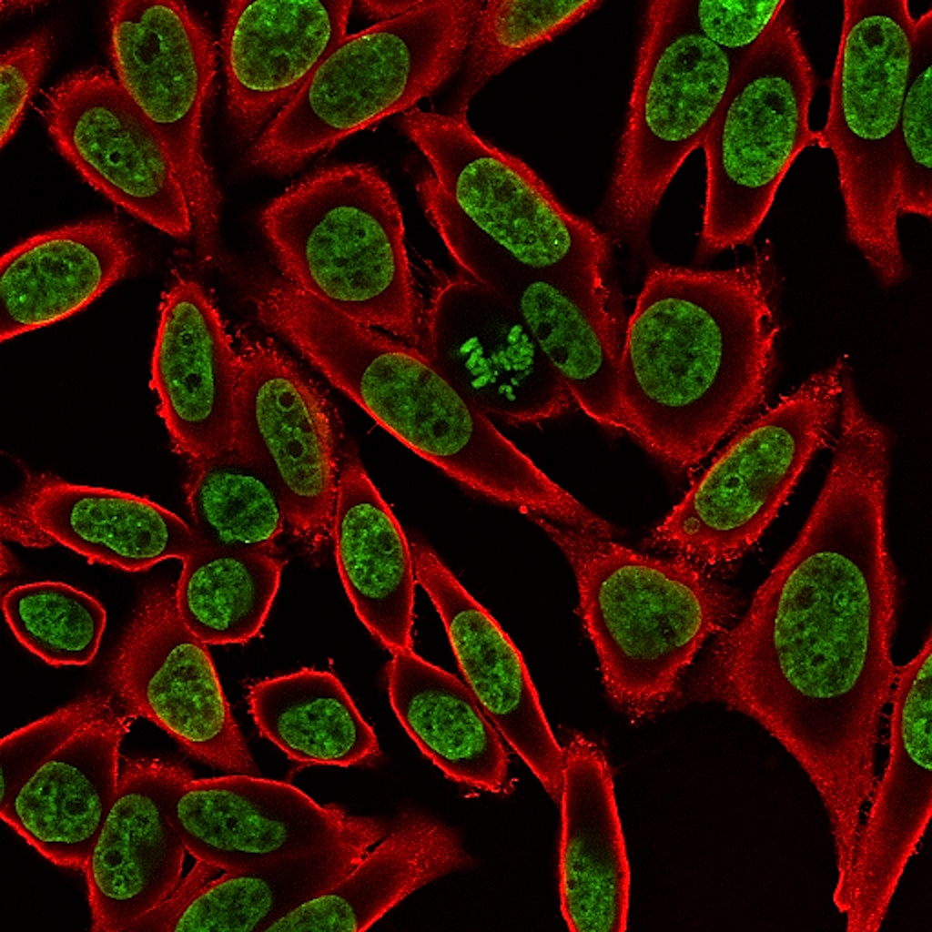
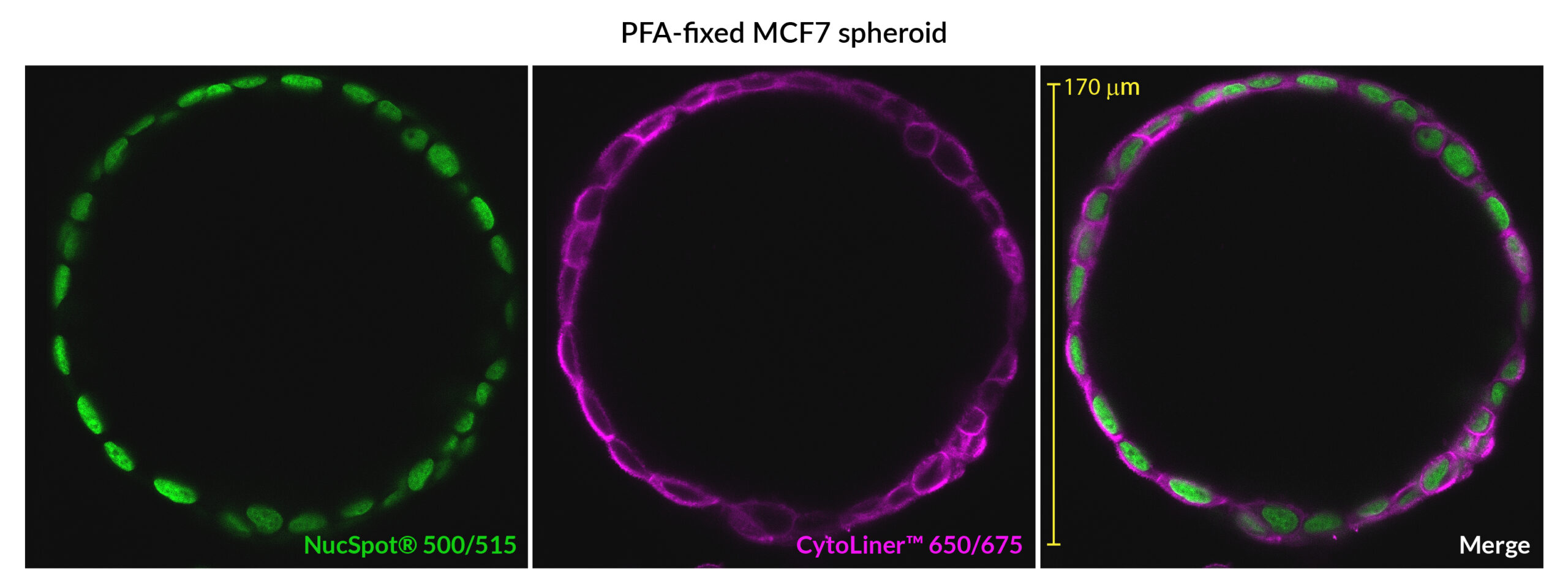
Bright, no-wash nuclear counterstains available from green to near-infrared. Designed for fixed-cell imaging and selective dead cell staining in live cultures without cytoplasmic background.

NucSpot® Nuclear Stains

Add to cart
SKU: 41040, 41033, 41036, 41037, 41034, 41035, 41038 Category: Tags: Brand:

Membrane-impermeant, nuclear-specific DNA counterstains for fixed cells and dead cell detection

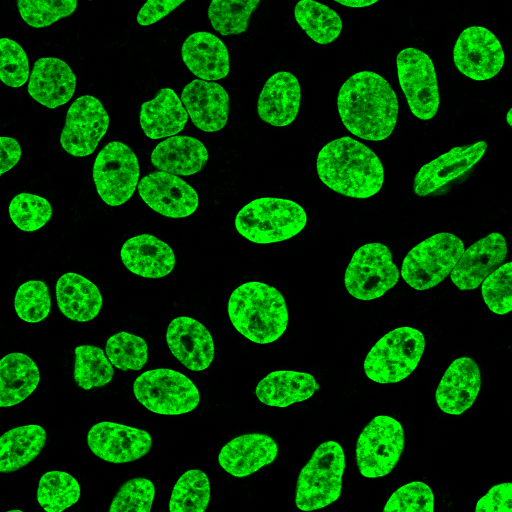
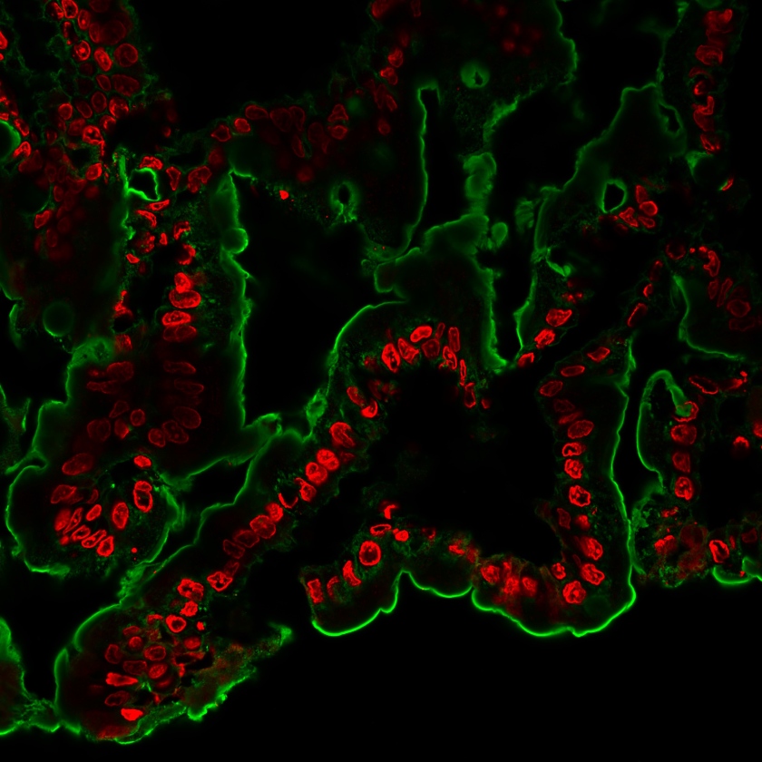
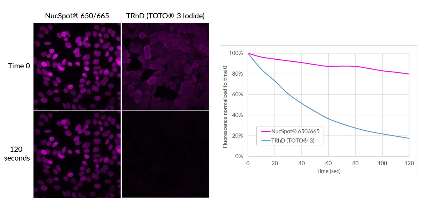
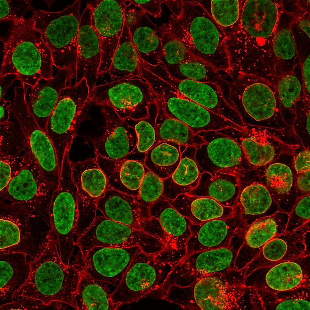
NucSpot® Nuclear Stains are cell membrane-impermeant, DNA-selective fluorescent dyes developed for clean, nuclear-specific counterstaining in fixed and permeabilized cells. Unlike traditional nuclear dyes such as DAPI or Hoechst that may undergo photoconversion or UV-induced channel cross-talk, NucSpot® stains provide stable fluorescence across a wide range of visible, far-red, and near-IR channels.

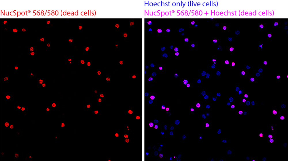
These dyes exhibit minimal fluorescence until bound to DNA, enabling no-wash workflows with reduced background. In live cultures, NucSpot® selectively stains dead or membrane-compromised cells, making them ideal for viability analysis by microscopy or flow cytometry.

Key Features

  • Nuclear-specific staining for fixed and permeabilized cells
  • Selective dead cell staining in live cultures
  • No wash required
  • Minimal fluorescence until DNA binding
  • Wide color selection from FITC to near-IR
  • Reduced photoconversion compared to UV-excited dyes
  • Compatible with fluorescence microscopy and flow cytometry

Why Choose NucSpot® Over Traditional Nuclear Stains?

Common blue nuclear dyes (DAPI, Hoechst) can photoconvert after UV exposure and cause cross-talk in other detection channels.

NucSpot® stains were developed to:

  • Eliminate UV-induced spectral interference
  • Provide brighter nuclear specificity without cytoplasmic staining
  • Avoid RNase treatment required by some nucleic acid dyes
  • Enable multiplex imaging in visible and far-red channels

Unlike PI, TOTO®, TO-PRO®, and similar dyes that stain both nucleus and cytoplasm, NucSpot® stains selectively label nuclear DNA in fixed cells.
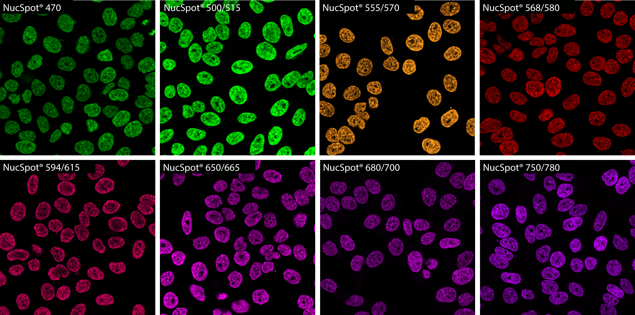
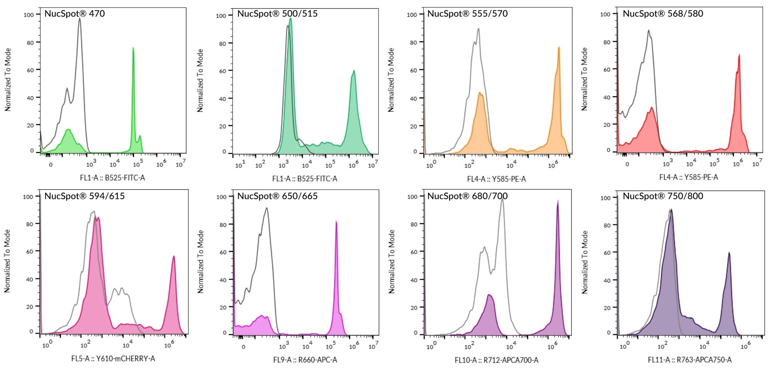
PFA-fixed, Triton® X-100 permeabilized HeLa cells stained with NucSpot® Nuclear Stains in PBS and imaged by confocal microscopy without a wash step. All stains were used at 1X concentration except for NucSpot® 750/780, which was used at 5X concentration in order to image using 640 nm excitation.
Live dead discrimination by flow cytometry using NucSpot® Nuclear Stains. A mixture of untreated and heat-killed Jurkat cells was stained with each NucSpot® Nuclear Stain and analyzed by flow cytometry on a BD CytoFLEX flow cytometerin the detection channel shown on the histogram x-axis. Solid peaks represent stained cells; open gray peaks represent unstained cells. The concentrations of each dye used were: 1X NucSpot® 470, 0.1X NucSpot® 500/515, 0.5X NucSpot® 555/565, 1X NucSpot® 568/580; 0.5X NucSpot® 594/615, 0.05X NucSpot® 650/665, 0.2X NucSpot® 680/700, 0.5X NucSpot® 750/780.
Photostability of NucSpot® 650/665 compared to Thiazole Red Homodimer. PFA-fixed, permeabilized HeLa cells were stained with 1X NucSpot® 650/665 or 200 nM Thiazole Red Homodimer (equivalent to TOTO®-3 Iodide). Cells were continuously imaged for 120 seconds by epifluorescence microscopy through a Cy®5 filter set, and images were captured every 10 seconds. Mean fluorescence normalized to time 0 for each image is plotted on the right. NucSpot® 650/665 fluorescence was well-preserved, while Thiazole Red/TOTO®3 fluorescence photobleached rapidly. In addition, NucSpot® 650/665 specifically stained the cell nucleus, while in the absence of RNase-pretreatment, TOTO®-3 stained both cytoplasm and nucleus.

Applications

  • Fluorescence microscopy
  • Flow cytometry
  • Live/dead discrimination
  • Long-term staining (24–72 hours)
  • Real-time imaging
  • Tissue staining
  • Multiplex immunofluorescence

Variations

Product Ex/Em (nm) Detection Channel Size Options SKU (Catalog No.)
NucSpot® 500/515 497/513 FITC* 20 µL, 100 µL 41040-T (20 µL), 41040 (100 µL)
NucSpot® 555/570 559/566 Cy®3 or PE* 20 µL, 100 µL 41033-T (20 µL), 41033 (100 µL)
NucSpot® 568/580 572/583 Cy®3 or PE* 20 µL, 100 µL 41036-T (20 µL), 41036 (100 µL)
NucSpot® 594/615 603/613 Texas Red® or PE-Texas Red®* 20 µL, 100 µL 41037-T (20 µL), 41037 (100 µL)
NucSpot® 650/665 653/671 Cy®5 or APC* 20 µL, 100 µL 41034-T (20 µL), 41034 (100 µL)
NucSpot® 680/700 683/707 Cy®5.5* 20 µL, 100 µL 41035-T (20 µL), 41035 (100 µL)
NucSpot® 750/780 757/780 Cy®7 or APC-Cy®7* 20 µL, 100 µL 41038-T (20 µL), 41038 (100 µL)

Special Highlights

NucSpot® 500/515

Improved alternative to NucSpot® 470. Stable in culture medium and suitable for multi-day live/dead staining.

NucSpot® 650/665

Superior nuclear specificity compared to first-generation far-red nuclear dyes such as RedDot™2 or Draq7™.

NucSpot® 680/700 & 750/780

Unique far-red and near-IR spectral options for multiplex and high-dimensional imaging panels.

NucSpot® Far-Red (Flow Cytometry Specific)

Designed as an improved alternative to 7-AAD with reduced PE-Texas Red® bleed-through. Optimised for flow cytometry DNA content and viability analysis.

Specification

Attribute Specification
Size 20 µL, 100 µL
Probe cellular localization Nucleus
For live or fixed cells For fixed cells
Assay type/options Live/dead discrimination, Long term staining (24–72h), No-wash staining, Real-time imaging, Tissue staining
Detection method/readout Fluorescence microscopy, Flow cytometry
Cell permeability Membrane impermeant
Apoptosis/viability marker Dead cell stain
Fixation options Fix before staining (formaldehyde), Fix before staining (methanol), Permeabilize before staining
Colors Green, Orange, Red, Far-red, Near-infrared
Storage Conditions Store at 2 to 8 °C

Documentation