Immunofluorescence (IF) imaging is a core technique for studying cardiomyocyte structure, function, and response to stimuli. As induced pluripotent stem cell (iPSC) derived cardiomyocytes become increasingly adopted for disease modelling, drug screening, and translational research, there is growing demand for robust plasma membrane staining strategies that preserve cellular integrity while enabling accurate imaging.
Plasma membrane IF imaging in cardiomyocytes presents unique challenges. These cells are sensitive to cytotoxic reagents, exhibit active membrane trafficking, and are often studied in advanced formats such as 3D organoids and co culture systems.
In this blog, we’ll explain why plasma membrane IF staining is both valuable and technically demanding, highlight common pitfalls, and discuss practical approaches to selecting appropriate membrane dyes and optimised staining strategies.
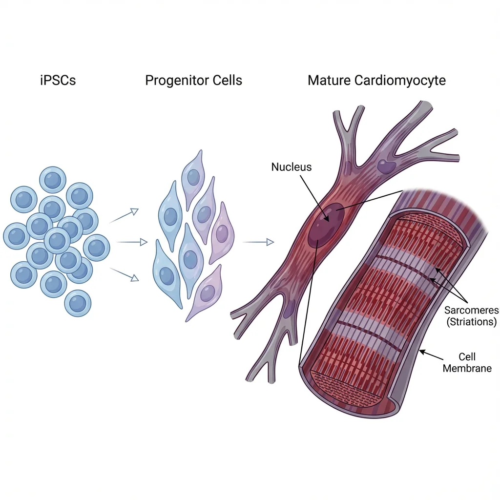
What Are iPSC derived cardiomyocytes?
Induced pluripotent stem cells (iPSCs) are somatic cells reprogrammed into a pluripotent state, a breakthrough developed by Dr Shinya Yamanaka, who was awarded the 2012 Nobel Prize in Physiology or Medicine for this discovery. iPSCs are now widely used in disease modelling, drug screening, and regenerative medicine, enabling the generation of specialised cell types such as insulin producing cells for diabetes and neurons for neurological disease. Their ability to provide patient specific, human relevant models has made iPSCs a cornerstone of modern translational research.
In cardiovascular research, iPSC derived cardiomyocytes (iPSC CMs) provide an experimentally accessible human platform for disease modelling, early stage human development studies, predictive toxicology, and therapeutic discovery. They recapitulate key structural and functional features of native heart cells, including organised sarcomeres, ion channel activity, and spontaneous contractility (Karakikes et al., 2015).
Characteristics of Cardiomyocytes Relevant to Imaging
Cardiomyocytes exhibit several features that directly impact plasma membrane immunofluorescence imaging:
High membrane sensitivity The sarcolemma hosts ion channels, transporters, and junctional proteins critical for electrophysiological function.
Contractile activity Spontaneous or paced beating introduces motion artefacts and increases susceptibility to membrane perturbation.
Low tolerance to cytotoxic reagents Cardiomyocytes are highly sensitive to dyes, solvents, and fixation conditions that may be well tolerated by other cell types.
Active membrane turnover Endocytosis and membrane recycling increase the risk of dye internalisation and signal redistribution.
These characteristics necessitate careful handling, gentle staining conditions, and the use of dyes with low cytotoxicity and high plasma membrane stability.
Why Plasma Membrane Imaging Matters in Cardiomyocytes Imaging
Plasma membrane immunofluorescence imaging plays a central role in cardiomyocyte studies by enabling:
Accurate cell boundary segmentation for quantitative imaging
Visualisation of sarcolemmal proteins such as ion channels and adhesion complexes
Spatial mapping of signalling events relative to the cell surface
Reliable identification of cardiomyocytes in co culture and mixed populations
Compared with nuclear based segmentation, plasma membrane imaging provides superior morphological definition in elongated, branched cardiomyocytes and densely packed syncytial cultures.
In advanced models such as 3D organoids and co-culture systems, plasma membrane staining improves the identification of individual cells within dense or mixed populations. Compared with nuclear-based segmentation, plasma membrane labelling provides improved morphological accuracy and supports quantitative imaging analyses.
Core Challenges in Plasma Membrane Immunofluorescence Staining
Despite its importance, plasma membrane immunofluorescence staining presents several technical challenges in cardiomyocyte imaging workflows:
1. Accessibility and Antibody Penetration
Surface epitopes can be masked by the lipid bilayer or glycocalyx. Too little permeabilisation limits access, while over permeabilisation disrupts membrane localisation. In 3D organoids, diffusion limits can cause surface only or hollow staining.
2. Dye Internalisation and Loss of Surface Signal
During live cell staining, membrane turnover drives internalisation into intracellular compartments, reducing boundary contrast. Uptake varies with dye chemistry, and can be mitigated by reduced temperature staining, fixation before labelling, or choosing dyes with longer surface retention.
3. Dye Detachment and Cell-to-Cell Transfer
In dense co-culture systems and cardiomyocyte syncytia, detached dyes can transfer between neighbouring cells, confounding cell identity and segmentation. Stable membrane association is critical for multiplexing and tracking.
4. Limited Surface Target Availability
Cell surface targets may be low abundance or masked by glycosylation or extracellular matrix, contributing to weak or uneven plasma membrane immunofluorescence staining.
5. Functional Perturbation of Cells
Membrane labelling can alter membrane properties or trigger clustering and signalling changes. This is particularly relevant in cardiomyocytes, where the sarcolemma supports ion exchange and membrane labels can affect action potentials or contractile beating.
6. Assay-Dependent Bias
Fixation, permeabilisation, and lipid extraction can change membrane composition and apparent localisation, and assays probing secretion or turnover can further bias staining patterns across fixed cell staining workflows.
Choosing the Right Plasma Membrane Dye
Selecting an appropriate plasma membrane dye is critical when working with sensitive cells such as iPSC-derived cardiomyocytes in immunofluorescence workflows. An effective plasma membrane dye should offer:
Low cytotoxicity
High plasma membrane stability
Reduced internalisation with sustained surface signal
Compatibility with fixation and permeabilisation for fixed cell staining
Clear cell boundary definition for imaging and segmentation
Suitable spectral options from blue to near-IR
Biotium offers a robust suite of membrane stains tailored for different workflows, from live cell imaging to IF compatible plasma membrane labelling. Use the guide below to select the best dye for your experiments:
Built for formaldehyde fixed cells with mild permeabilisation; reliable boundary staining that stays compatible with antibody staining; colours blue to near-IR
Covalent labelling of cell surface proteins; wide colour selection; performs well with fixation and permeabilisation for consistent boundaries
Practical Tips for Success
Plan stain order wisely: For co-staining with antibodies, start with membrane staining (especially with CytoLiner™) before blocking and antibody incubations.
Optimise permeabilisation: Adjust detergent concentration and incubation time based on your cell type and structure.
Use blockers: Fish gelatin or serum can help reduce non-specific background from complex tissues.
Choose colours carefully: Stay mindful of emission overlap when designing multi-colour assays.
Control temperature during live staining: Performing membrane labelling at reduced temperatures can slow endocytic uptake and improve surface signal retention in highly endocytic cells.
Match fixation to membrane preservation: Paraformaldehyde generally maintains membrane architecture, while methanol can extract lipids and disrupt surface staining unless labelling is performed prior to fixation.
Time imaging in contractile models: In beating iPSC-derived cardiomyocytes, allow sufficient recovery after staining or fixation and optimise acquisition timing to reduce motion-related artefacts.
Conclusion
Plasma membrane IF staining is an invaluable tool for modern cell biology — but its success hinges on choosing the right dyes, matching fixation/permeabilisation strategies, and understanding the biological context of your cells. Whether you’re outlining the sarcolemma of iPSC-derived cardiomyocytes, mapping cellular interactions in co-culture, or visualising boundaries in 3D organoids, a thoughtful stain and workflow selection makes all the difference.
Reference:
Hassdenteufel, S., & Schuldiner, M. (2021). Show your true color: Mammalian cell surface staining for tracking cellular identity in multiplexing and beyond. Current Opinion in Chemical Biology, 66, 102102. https://doi.org/10.1016/j.cbpa.2021.102102
Karakikes, I., Ameen, M., Termglinchan, V., & Wu, J. C. (2015). Human induced Pluripotent Stem Cell–Derived cardiomyocytes. Circulation Research, 117(1), 80–88. https://doi.org/10.1161/circresaha.117.305365
Discover the top 10 biotechnology trends shaping 2026, from in vivo editing and RNA therapeutics to spatial omics and regenerative medicine innovation.
Summary The field of cancer therapy is undergoing transformative changes, driven by the following trends: iPSC-derived MSCs, MSCs-derived EVs, and Cas9-mediated gene editing in MSCs.
HOW CAN WE HELP YOU?Our specialists are to help you find the best product for your application. We will be happy to help you find the right product for the job.