

$357.00
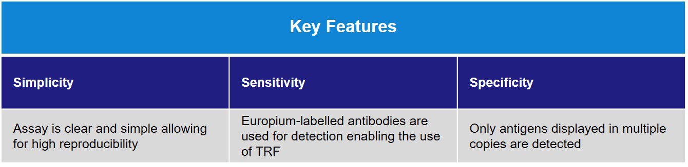
The TRIFic™ exosome detection assay delivers clear and consistent data quickly by providing quantitative data from purified or unpurified samples, including direct measurement of exosomes in plasma in a convenient 96-well format. It is similar to an ELISA assay, but the target is detected directly with a Europium label without any enzymatic reaction. TRIFic™ exosome detection assays are being made available for widely used exosome markers, allowing for identification of CD9+/CD81+/CD63+ – EVs.
| Code | Description |
|---|---|
| EX101 | TRIFic™ CD9 Exosome Assay, 96 wells |
| EX102 | TRIFic™ CD63 Exosome Assay, 96 wells |
| EX103 | TRIFic™ CD81 Exosome Assay, 96 wells |
| EX-P31 | TRIFic™ CD81 Exosome Assay, 96 wells |
Which Exo-spin™ kit shall I choose for my sample volume?
Assay principle
Exosomes typically have multiple copies of tetraspanin facing towards the attachment surface and additional marker molecules available for detection. Based on this property, the assay consists of a monoclonal antibody (labeled with biotin) bound to a streptavidin-coated plate which captures protein present on the surface of exosomes (see below). Subsequently, an identical monoclonal antibody (labeled with europium) is used for detection.
Exosomes provide an ideal structure to link CD9, CD63 or CD81 molecules and allow detection of the tetraspanin of interest. Using a europium fluorophore provides high levels of sensitivity for the assay, which is able to detect small changes in the abundance of the target tetraspanin protein even within unpurified complex biological samples, such as blood plasma and cerebrospinal fluid (CSF).
Protocol Overview
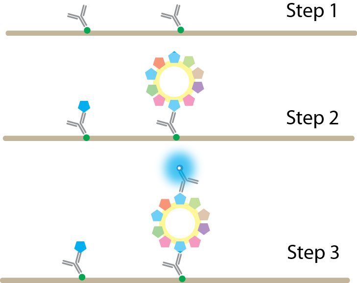 Step 1. Biotinylated antibody is bound to the streptavidin-coated assay plate.
Step 2. Biological samples containing exosomes are added to the plate. Exosomes bearing the appropriate antigen (CD9, CD63, or CD81) and any free antigen are captured by the immobilized antibody.
Step 3. Europium-labeled antibody is added and binds specifically to exosome antigens. Free antigen is not bound by the europium-labeled antibody (since the epitope is already occupied by the immobilized antibody) and is therefore not detected. Samples are read in a time-resolved fluorescence plate reader.
Fluorophores
Fluorophores are chemical substances that emit light following excitation by light or other electromagnetic radiation. The emission of light from a fluorophore is maximal immediately following excitation and decays over a period of time. Time-resolved fluorimetry uses fluorophores which have long decay periods. For such fluorophores, measurement of emitted light can be performed when the excitation light is no longer present, thus increasing sensitivity.
Europium
Europium is a fluorophore which produces an extended emission decay and has a wide Stokes shift with maximal excitation at 340 nm and peak emission at 615 nm. TRIFic™ assays are time-resolved immunofluorescence assays which utilize europium and have been developed to measure the abundance of CD9, CD63, or CD81 protein specifically associated with exosomes in biological fluids including urine, saliva, cell culture medium, cerebrospinal fluid (CSF), and blood plasma.
Applications
The TRIFic™ detection assay is ideal for detection of specific markers of exosomes, for purified or unpurified samples.
[accordions]
[accordion title=”Product Data”]
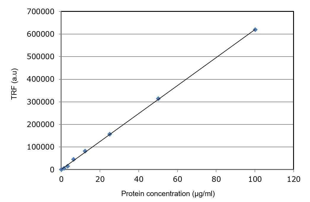
Titration of purified exosomes using TRIFic™ exosome assay shows a linear relationship between quantity of exosomes and TRF units (time-resolved fluorescence arbitrary units) over a wide range. The assay is able to detect sub-microgram levels of exosomes.
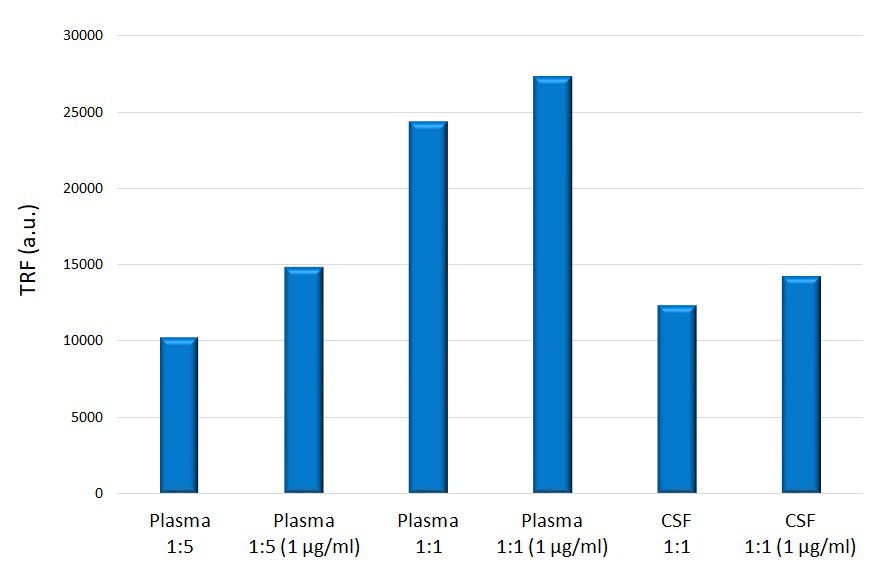
Biological samples (diluted 1:1 or 1:5 with PBS) spiked with purified exosomes demonstrates the sensitivity of the assay even in complex biological fluids such as plasma and cerebrospinal fluid.
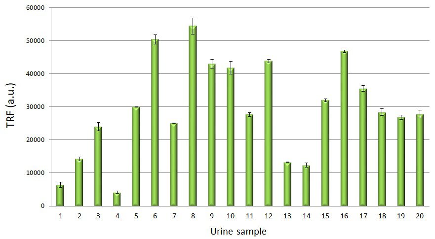
TRIFic™ exosome analysis of CD9 in 20 cancer patient urine samples demonstrating a wide range of exosomal content.
TRIFic™ exosome detection assay scientific data on unpurified samples
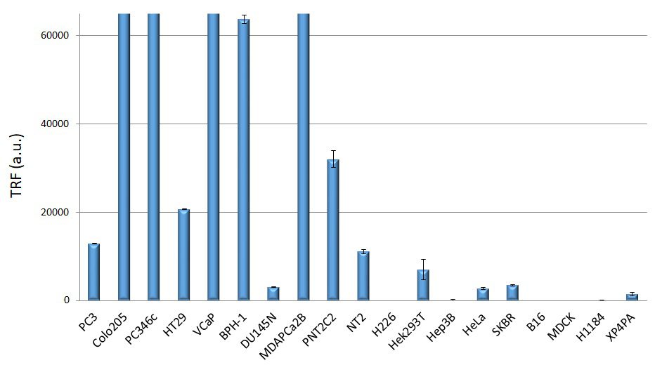
CD9 TRIFic™ exosome assay performed for a variety of cell lines (n=2). Over 80 samples plus exosome controls can be tested with a single kit.
Exosome marker detection for cancer cell lines
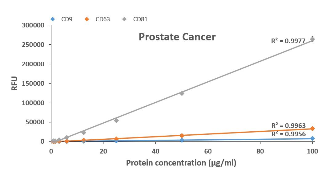
Marker detection on exosomes from prostate cancer cells using TRIFic™. Each datapoint represents an average (n=3).
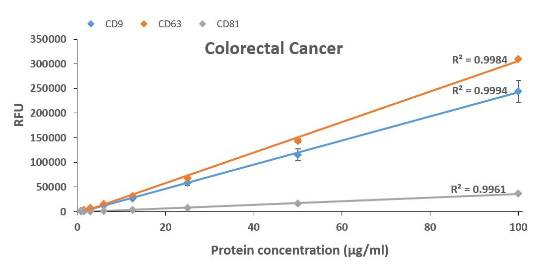
Marker detection on exosomes from colorectal cancer cells using TRIFic™. Each datapoint represents an average (n=3).
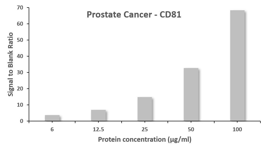
Signal to blank ratio of CD81 detection on exosomes from prostate cancer cells. To obtain the datapoints average values were used.
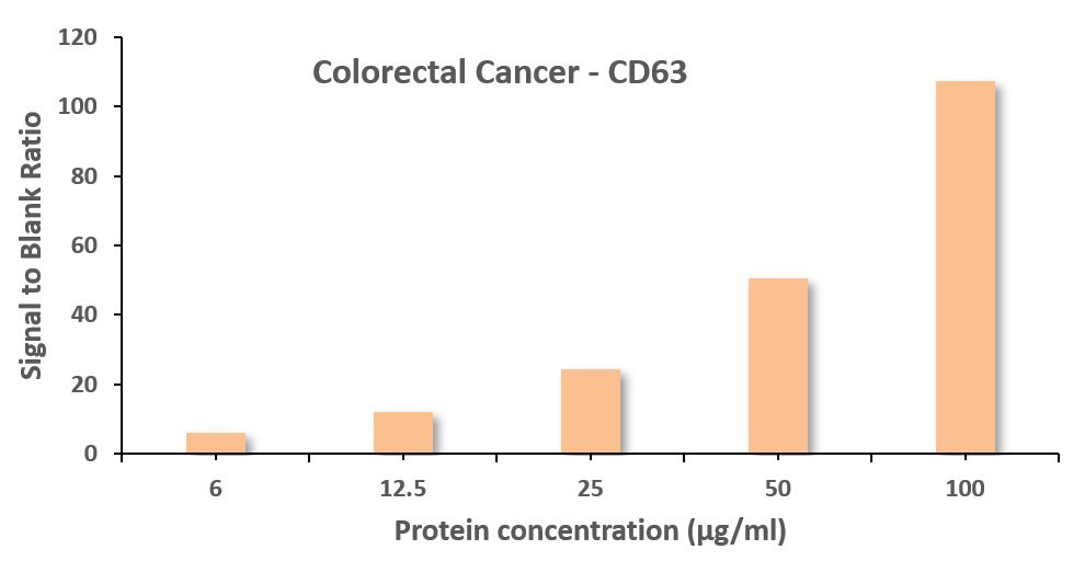
Signal to blank ratio of CD81 detection on exosomes from colorectal cancer cells. To obtain the datapoints average values were used.
[/accordion]
[accordion title=”User guide”] EX101_CD9_TRIFic_User_Guide. CD63__clone_CGS73B__Datasheet. CD81__clone_CGS36K__Datasheet[/accordion]
[accordion title=”MSDS/Tech Sheet”] TRIFic_kits_MSDS[/accordion]
[accordion title=”Product Brochure”] CellGS exosome brochure.pdf[/accordion]
[/accordions]
Contact our Customer Care, Sales & Scientific Assistance
Consult and asked questions about our products & services
Documentation of Technical & Safety Data Sheet, Guides and more..
We gladly support you by keeping you updated on our latest products and the developments around our services.
362 Upper Paya Lebar Rd, #07-15,
Singapore 534963
WeChat ID: +65 8608 0974
© 2015-2023 Atlantis Bioscience Pte Ltd. All rights reserved. Co Reg No: 201539516N