PRODUCT ATTRIBUTES
| Cellular localization | Mitochondria |
|---|---|
| For live or fixed cells | For live/intact cells |
| Assay type/options | No-wash staining, Real-time imaging |
| Cell permeability | Membrane permeant |
| Potential dependence | Mitochondrial potential dependent, Mitochondrial potential-independent |
| Apoptosis/viability marker | Mitochondrial potential |
| Fixation options | Fix before staining (formaldehyde) |
| Application notes | Cells can be fixed with formaldehyde before staining with MitoView™ Green only, Mitochondrial localization of MitoView™ Blue and MitoView™ 720 is potential-dependent, MitoView™ 633 fluorescence is mitochondrial potential-dependent, MitoView™ Green staining is mitochondrial potential-independent |
| Colors | Blue, Green, Far-red, Near-infrared |
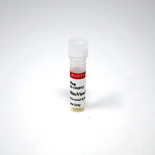
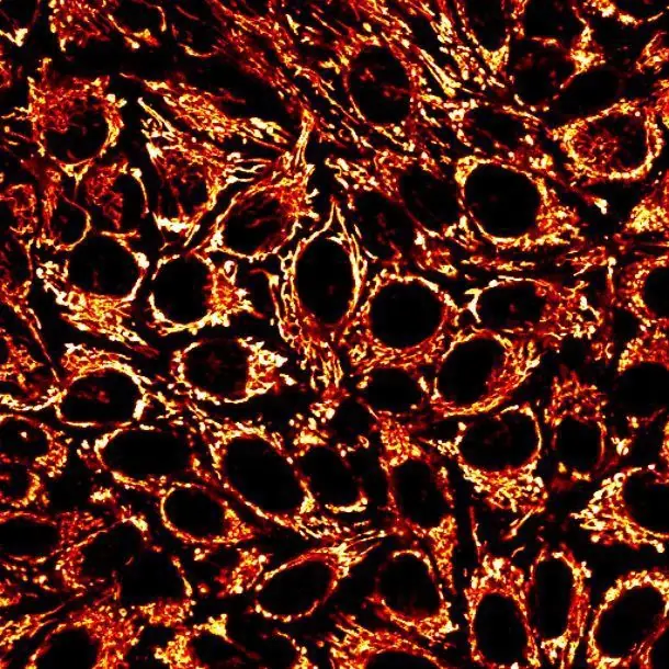
MitoView™ dyes are fluorogenic mitochondrial stains for live cells. The dyes rapidly accumulate in mitochondria and can be imaged without washing. They are available with blue, green, far-red, and near-infrared fluorescence. Cells can be fixed before staining with MitoView™ Green only, the other MitoView™ dyes are for use in live cells only.
Features
Note: For staining mitochondria in fixed cells or tissue sections, we recommend using one our Mitochondrial Marker Antibodies, available with a wide selection of bright and photostable CF® dyes and other conjugations.
MitoView™ dyes can also be used to stain mitochondria in yeast and bacteria (gram-positive and gram-negative). See our Cellular Stains Table for more information on how our dyes stain various organisms.
MitoView™ Dyes
| Product | Abs/Em | Detection channel | Potential-dependent? | Catalog no. | Size |
|---|---|---|---|---|---|
| MitoView™ 405 | 398/440 nm | DAPI | Partial† | 70070-T | 50 ug |
| 70070 | 20×50 ug | ||||
| MitoView™ Green | 490/523 nm | FITC, GFP | No | 70054-T | 50 ug |
| 70054 | 20×50 ug | ||||
| MitoView™ 633 | 622/648 nm* | Cy®5, APC* | Yes | 70055-T | 50 ug |
| 70055 | 20×50 ug | ||||
| MitoView™ 650 | 644/670 nm | Cy®5, APC* | Partial† | 70075-50ug | 50 ug |
| 70075 | 20×50 ug | ||||
| MitoView™ 720 | 720/758 nm** | Cy®5, Cy®7** | Partial† | 70068-T | 50 ug |
| 70068 | 20×50 ug |
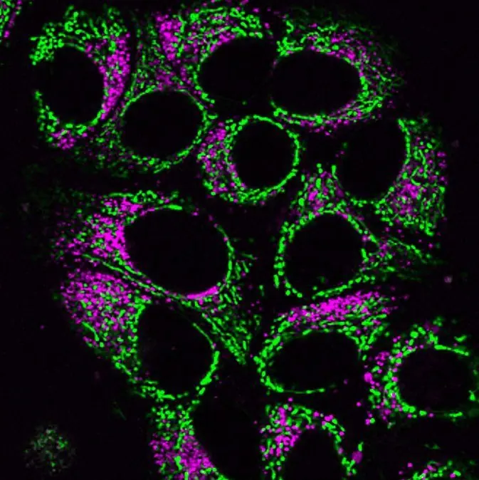
* MitoView™ 633 also has red fluorescence in the Cy®3 channel and is not recommended for use with other red dyes.
**While optimal for Cy®7 settings, MitoView™ 720 is bright enough to be imaged in the Cy®5 channel.
† Dyes localize to the cytoplasm after mitochondrial depolarization, but still retain fluorescence.MitoView™ 405
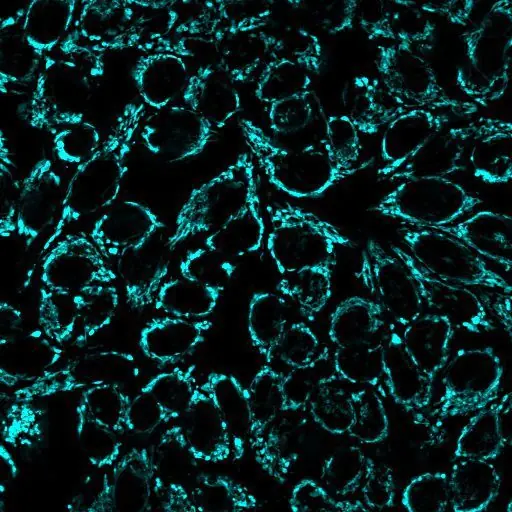
MitoView™ 405 is a blue fluorescent mitochondrial dye with absorbance/emission at 398/440 nm, suitable for detection by confocal microscopy or flow cytometry using settings for DAPI or Pacific Blue®. The dye is membrane permeable and becomes brightly fluorescent upon accumulation in the mitochondrial membrane. Mitochondrial localization is dependent on mitochondrial membrane potential; when membrane potential is disrupted the dye relocalizes to the cytoplasm, but still retains fluorescence. The dye is designed for use in live cells and is not fixable. MitoView™ 405 is a replacement for our original MitoView™ Blue, and has improved photostability.
MitoView™ Green
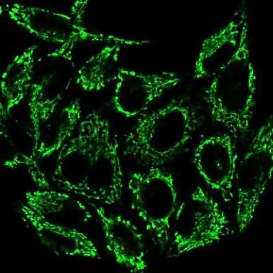
MitoView™ Green is a green fluorescent mitochondrial dye with properties similar to those of MitoTracker® Green FM. The dye is non-fluorescent until it partitions into the mitochondrial membrane. The staining relies on mitochondrial mass, not on mitochondria membrane potential. Thus, the dye can be used to stain mitochondria in both live cells and fixed cells.* MitoView™ Green is spectrally similar to FITC.
*Note: For optimal staining of mitochondria in fixed cells or tissue sections, we recommend using one our Mitochondrial Marker Antibodies, available with a wide selection of bright and photostable CF® dyes and other conjugations.
Note: MitoView™ Green may be somewhat membrane potential-dependent in yeast cells.
MitoView™ 633
MitoView™ 633 is a far-red fluorescent mitochondrial dye with absorbance/emission at 622/648 nm. The dye is membrane permeable and becomes brightly fluorescent upon accumulation in the mitochondrial membrane. Staining is dependent on mitochondrial membrane potential, and can be used to monitor mitochondrial membrane potential in intact cells. The dye is designed for use in live cells, and is not fixable. Note: The optimal detection settings for MitoView™ 633 are the same as for Cy®5 and other far-red dyes. However, the dye also has visible red fluorescence and can be imaged using Cy®3 settings as well. As a consequence, the dye cannot be used for two-color imaging with other red probes.
MitoView™ 650
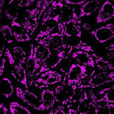
MitoView™ 650 is a far-red fluorescent mitochondrial dye that is not dependent on mitochondrial membrane potential. The dye has excitation/emission at 644/670 nm for the Cy®5 channel. Unlike MitoView™ 633, it does not bleed into the visible red channel, and so can be combined with red probes for multicolor imaging. The dye fluorescence is not lost after mitochondrial depolarization or fixation, but localization becomes non-specific.
MitoView™ 720
MitoView™ 720 is a near-infrared mitochondrial dye with absorbance/emission at 720/758 nm. While optimally detected using Cy®7 settings, the dye is bright enough to be imaged in the Cy®5 channel, and can be combined with visible red fluorescent probes. Mitochondrial localization is dependent on mitochondrial membrane potential; when membrane potential is disrupted the dye relocalizes to the cytoplasm but still retains fluorescence.
In addition to MitoView™ dyes, Biotium also offers several classical dyes for measuring mitochondrial membrane potential. These include JC-1, a ratiometric dye that forms red aggregates in healthy cells, which are reduced to the green monomeric state when mitochondrial membrane potential is lost. We also offer TMRM and TMRE, two red fluorescent dyes that are used to quantitatively measure mitochondrial membrane potential using the Nernst equation.
To view our wide selection of other cellular stains, visit our Cellular Stains technology page or see our Cellular Stains Brochure.
Note: MitoView™ Blue (70052) has been discontinued and replaced by the improved MitoView™ 405. The original MitoView™ Blue will be available for purchase as a special order while supplies last. Contact [email protected] to inquire about availability.
Protocols
SDS
Supporting Documents
Contact our Customer Care, Sales & Scientific Assistance
Consult and asked questions about our products & services
Documentation of Technical & Safety Data Sheet, Guides and more...
We gladly support you by keeping you updated on our latest products and the developments around our services.
362 Upper Paya Lebar Rd, #07-15,
Singapore 534963
© 2015-2023 Atlantis Bioscience Pte Ltd. All rights reserved. Co Reg No: 201539516N