🔥 Don’t Miss Out on our Blind Box Giveaway with a minimum spend of $280! 🔥 Don’t Miss Out on our Blind Box Giveaway with a minimum spend of $280! 🔥 Don’t Miss Out on our Blind Box Giveaway with a minimum spend of $280!
Add friend

ExoBrite™ WGA EV Staining Kits

ExoBrite™ WGA EV Staining Kits

ExoBrite™ WGA EV Staining Kits

ExoBrite™ WGA EV Staining Kits provide bright, low-background staining of purified and bead-bound EVs, offering broad compatibility across multiple sources.

ExoBrite™ WGA EV Staining Kits cover image

Add to cart
SKU: 30123-T, 30123, 30124-T, 30124, 30125-T, 30125, 30126-T, 30126 Categories: Tags: Brand:

Sensitive Stains for Purified or Bead-Bound EVs with Broad Compatibility

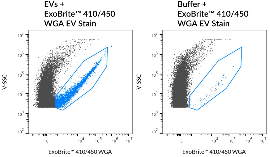
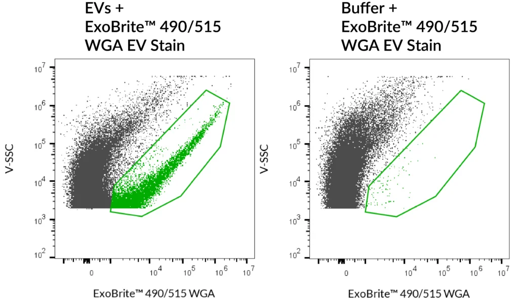
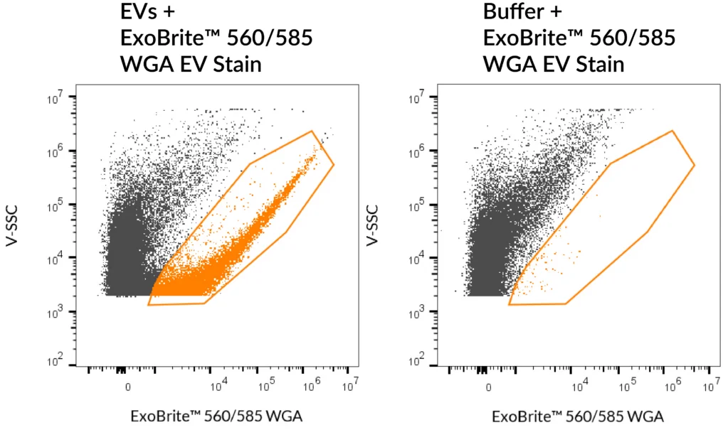
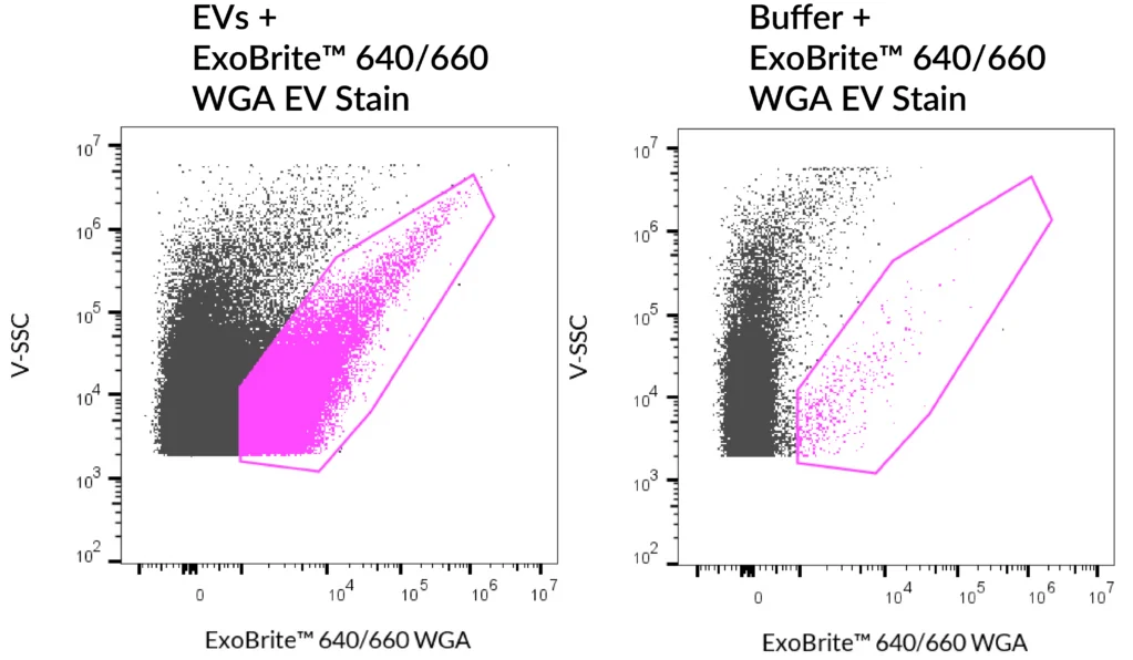
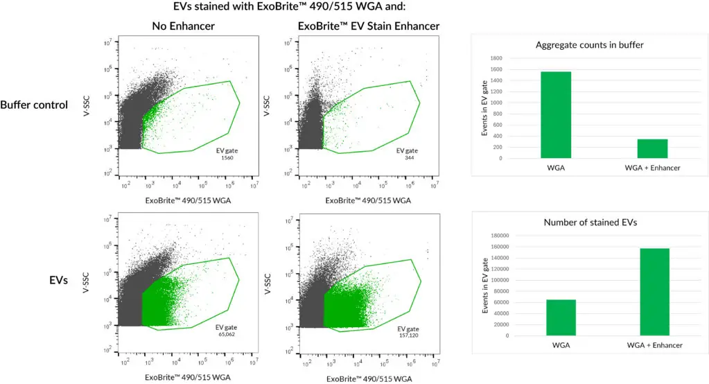
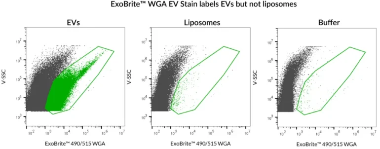
ExoBrite™ WGA EV Staining Kits are fluorescent conjugates of wheat germ agglutinin (WGA), specifically optimised for bright and sensitive staining of extracellular vesicles (EVs). Unlike hydrophobic membrane dyes that can aggregate or bind non-specifically, ExoBrite™ WGA EV Stains are formulated for clear, low-background staining in flow cytometry.

Validated on EVs derived from 9 cell lines, these stains are compatible with both purified EVs and bead-bound EVs, offering broad coverage across different EV sources. Each kit includes the ExoBrite™ WGA EV Stain and 1X PBS solution, ensuring convenient and reliable experimental use.

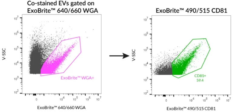
By binding to glycoproteins on EV membranes, ExoBrite™ WGA stains provide bright signal intensity and are fully compatible with antibody co-staining (e.g. CD9, CD63, CD81), enabling multi-parameter EV analysis.

Key Features

  • Optimally formulated WGA conjugates for EV staining
  • Compatible with both purified and bead-bound EVs
  • Bright signal, low background, ideal for flow cytometry
  • Broad compatibility – strong staining across 9 validated EV sources
  • Compatible with antibody co-staining for multiplex analysis
  • Available in four colours


SEC-purified EVs were stained with ExoBrite™ WGA EV Stains. MCF-7 derived EVs were stained with ExoBrite™ 410/450, ExoBrite™ 490/515, or ExoBrite™ 560/585. CHO derived EVs were stained with ExoBrite™ 640/660. 410/450 was detected in the Pacific Blue channel, 490/515 was detected in the FITC channel, 560/585 was detected in the PE channel, and 640/660 was detected in the APC channel.


ExoBrite™ 490/515 WGA EV Stain was used to stain either SEC-purified, Jurkat-derived EVs, or artificial liposomes. Robust staining was seen with the EVs, but the liposomes were not stained. The liposomes in this experiment were prepared from Presome® ACD-1, and were verified to be of similar size and concentration to the EVs using the lipophilic dye di-8-ANNEPS (data not shown). Analysis was performed on a CytoFLEX flow cytometer with SSC detected from the 405 nm laser, and ExoBrite™ detected in the FITC channel. Presome® is a registered trademark of NIPPON FINE CHEMICAL CO., LTD.


SEC-purified MCF-7-derived EVs were stained first with ExoBrite™ 490/515 CD81 in 100 uL, followed by 1X ExoBrite™ 640/660 WGA EV Stain. EVs were detected on a CytoFLEX LX flow cytometer in the FITC and APC channels. When we gated on ExoBrite™ 640/660 WGA-positive particles, ~60% were also positive for CD81.


Kit Components

  • ExoBrite™ WGA EV Stain
  • ExoBrite™ 1X PBS Solution


Notes

  • ExoBrite™ WGA EV Stains were validated on 9 cell line-derived EVs but may not stain EVs from all sources.
  • For ExoBrite™ 490/515 dye, a biotin blocking step is recommended when using streptavidin-coated beads or surfaces.
  • Biotium products are available only in Singapore and Thailand.


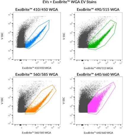
Explore ExoBrite™ WGA EV Staining Kits today – four colours are ready for your EV research.

*Please leave us a message during checkout to indicate which kit you need, and our team will process your order accordingly.

Variations

Product Ex/Em (nm) Detection Channels Size Options Catalog Number
ExoBrite™ 410/450 WGA EV Staining Kit 416/452 nm Pacific Blue™ 100 Labelings / 500 Labelings 30123-T / 30123
ExoBrite™ 490/515 WGA EV Staining Kit 490/516 nm FITC 100 Labelings / 500 Labelings 30124-T / 30124
ExoBrite™ 560/585 WGA EV Staining Kit 562/584 nm PE 100 Labelings / 500 Labelings 30125-T / 30125
ExoBrite™ 640/660 WGA EV Staining Kit 642/663 nm APC 100 Labelings / 500 Labelings 30126-T / 30126

Documentation