ExoBrite™ Streptavidin Magnetic Beads

ExoBrite™ Streptavidin Magnetic Beads

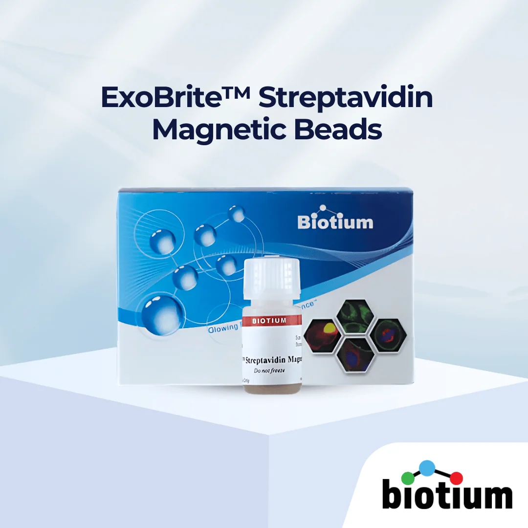
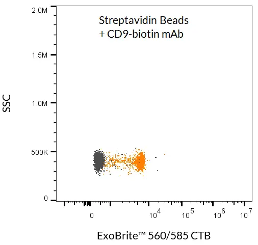
ExoBrite™ Streptavidin Magnetic Beads enable sensitive EV isolation with biotinylated antibodies, offering lower background and faster workflows.

ExoBrite™ Streptavidin Magnetic Beads cover image

Add to cart

ExoBrite™ Streptavidin Magnetic Beads

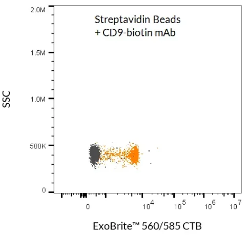
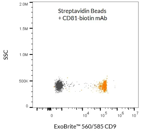
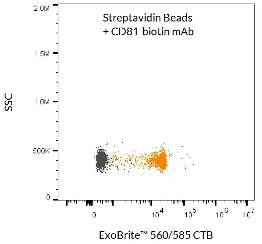
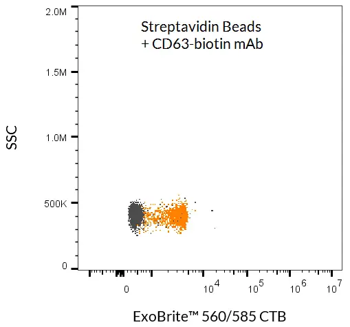
ExoBrite™ Streptavidin Magnetic Beads are streptavidin-coated magnetic polystyrene beads (4.5 µm diameter) validated for the isolation of extracellular vesicles (EVs) and exosomes. When combined with a biotinylated antibody (not included), these beads provide a simple, efficient method for EV capture from cell culture medium or biological fluids without the need for an overnight precipitation step.

Designed for lower background and higher sensitivity compared to conventional methods, ExoBrite™ beads enable reliable EV isolation followed by downstream analysis with flow cytometry, fluorescence microscopy, or western blot. Bead-bound EVs can also be lysed for protein or nucleic acid analysis, supporting multi-modal EV characterisation.

Key Features

  • Combine with a biotinylated antibody for EV capture
  • Isolate EVs from biological fluids without precipitation
  • Lower background, higher sensitivity than competitors
  • Magnetic-based separation for speed and convenience
  • Compatible with flow cytometry, microscopy, western blot, sequencing


Applications

  • EV/exosome isolation via antibody capture (CD9, CD63, CD81, or other membrane markers)
  • Detection of bead-bound EVs with antibody conjugates or fluorescent probes
  • Protein analysis by western blot
  • Nucleic acid profiling by sequencing


Notes: Biotium products are available only in Singapore and Thailand.


Streamline your EV workflows with ExoBrite™ Streptavidin Magnetic Beads – high-sensitivity isolation without precipitation.

*Please leave us a message during checkout to indicate which kit you need, and our team will process your order accordingly.

Documentation

Proven Performance

ExoBrite™ Streptavidin Magnetic Beads were used to isolate and detect EVs from tissue culture cell supernatant. The beads were first incubated with a biotinylated CD9 antibody, and then with concentrated MCF-7 cell supernatant. The EVs bound to the beads were stained with ExoBrite™ 560/585 CTB EV Stain and detected by flow cytometry in the PE channel (orange dots). Beads without antibody were also incubated with cell supernatant and stained, and no staining was seen (gray dots).
ExoBrite™ Streptavidin Magnetic Beads were used to isolate and detect EVs from tissue culture cell supernatant. The beads were first incubated with a biotinylated CD81 antibody, and then with concentrated MCF-7 cell supernatant. The EVs bound to the beads were stained with ExoBrite™ 560/585 CTB EV Stain and detected by flow cytometry in the PE channel (orange dots). Beads without antibody were also incubated with cell supernatant and stained, and no staining was seen (gray dots).

ExoBrite™ Streptavidin Magnetic Beads were used to isolate and detect EVs from tissue culture cell supernatant. The beads were first incubated with a biotinylated CD81 antibody, and then with concentrated MCF-7 cell supernatant. The EVs bound to the beads were stained with ExoBrite™ 560/585 CD9 Flow Antibody and detected by flow cytometry in the PE channel (orange dots). Beads without antibody were also incubated with cell supernatant and stained, and no staining was seen (gray dots).

ExoBrite™ Streptavidin Magnetic Beads were used to isolate and detect EVs from tissue culture cell supernatant. The beads were first incubated with a biotinylated CD63 antibody, and then with concentrated MCF-7 cell supernatant. The EVs bound to the beads were stained with ExoBrite™ 560/585 CTB EV Stain and detected by flow cytometry in the PE channel (orange dots). Beads without antibody were also incubated with cell supernatant and stained, and no staining was seen (gray dots).