🔥 Don’t Miss Out on our Blind Box Giveaway with a minimum spend of $280! 🔥 Don’t Miss Out on our Blind Box Giveaway with a minimum spend of $280! 🔥 Don’t Miss Out on our Blind Box Giveaway with a minimum spend of $280!
Add friend
ExoBrite™ CD9 Flow Antibody

ExoBrite™ CD9 Flow Antibody

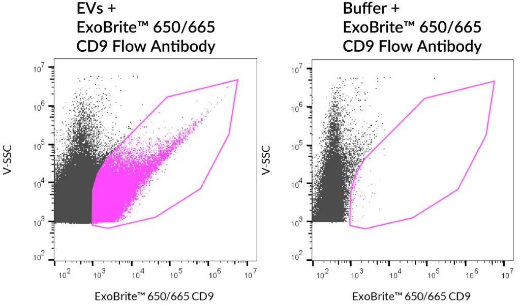
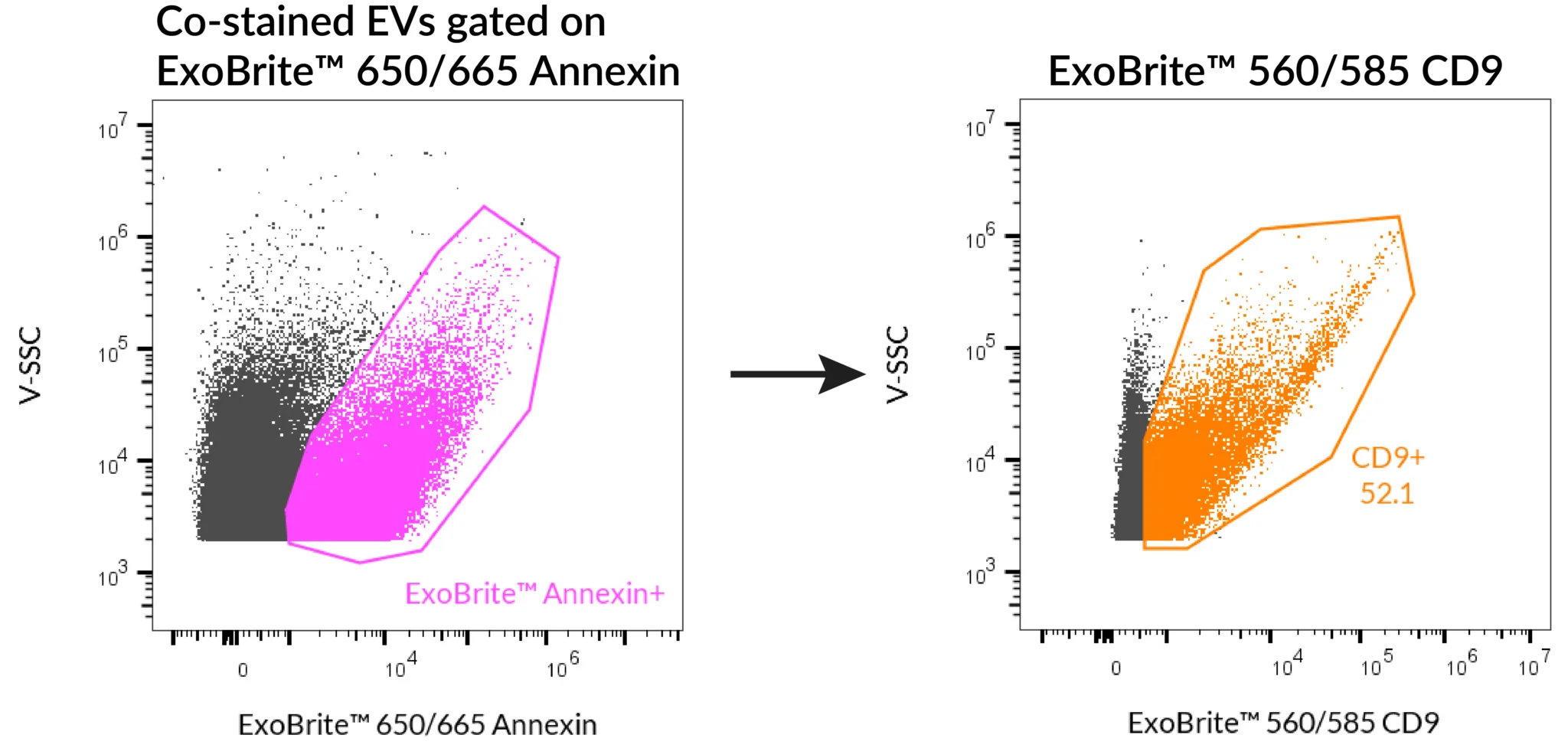
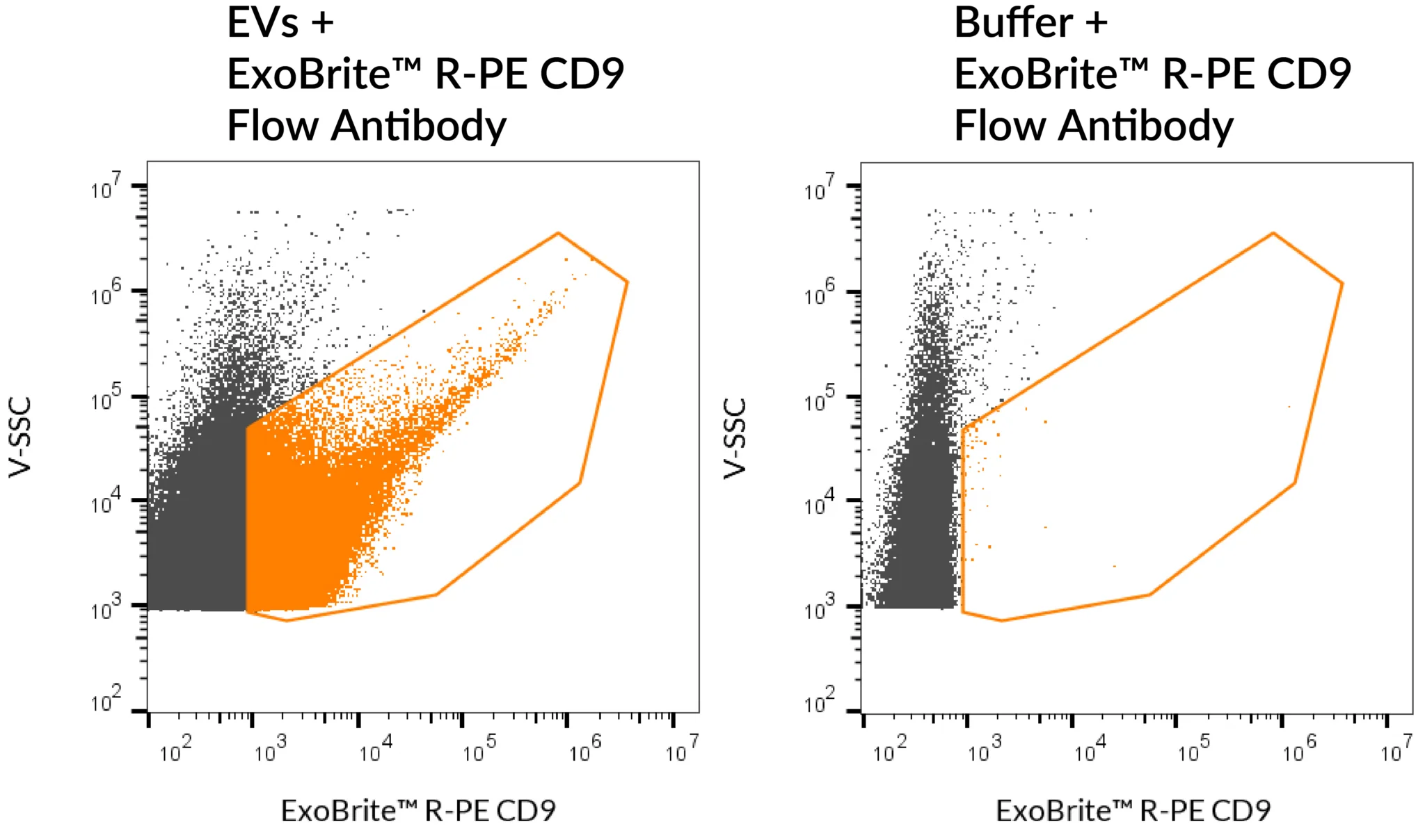
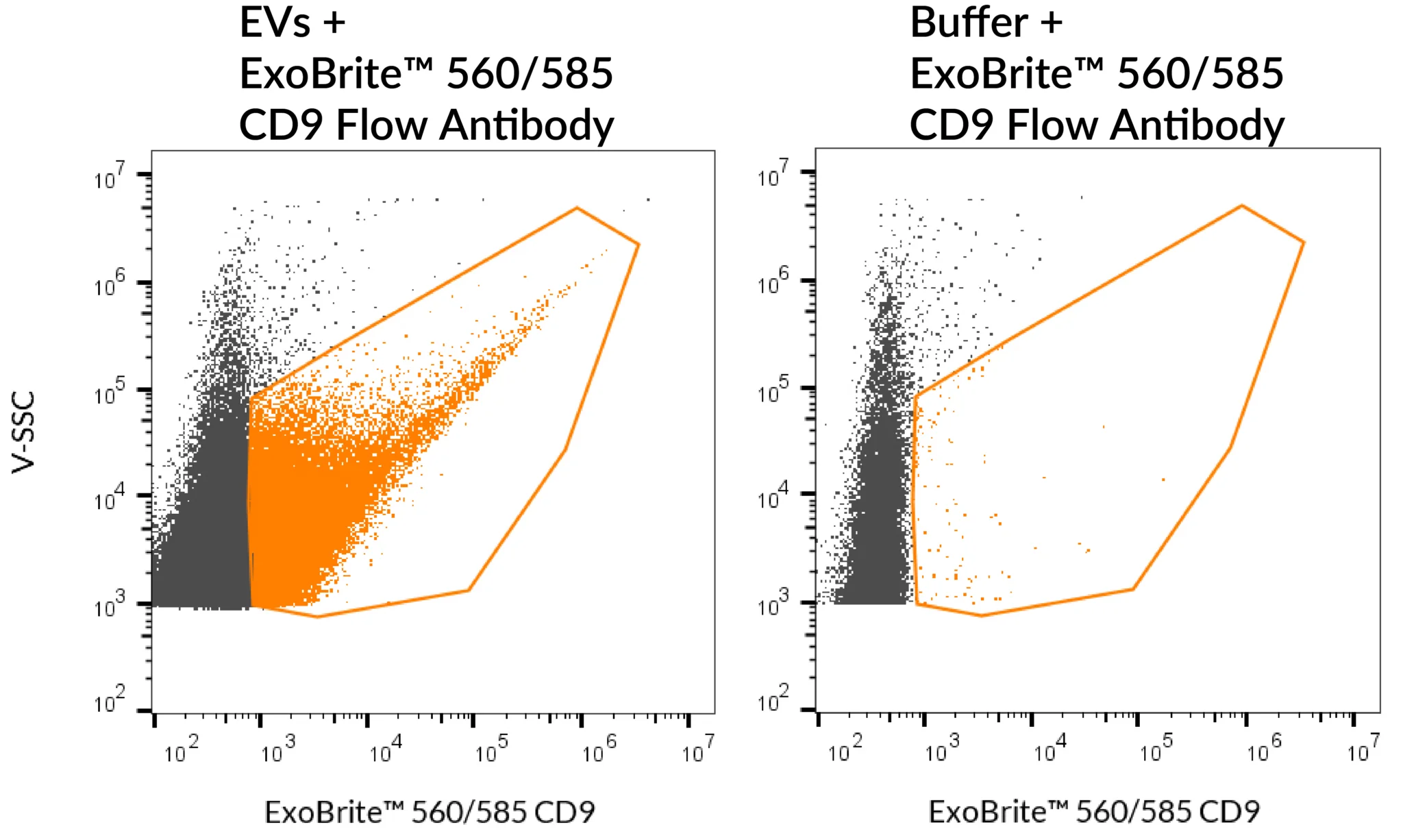
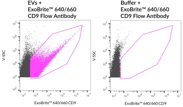
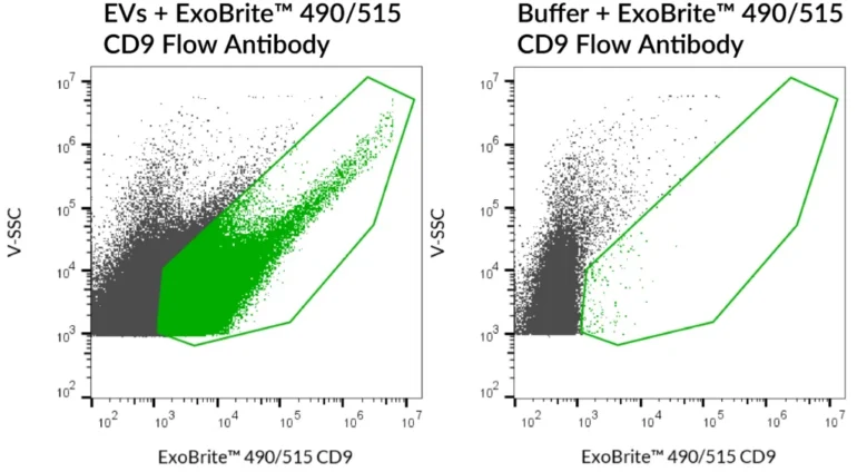
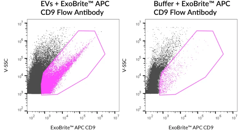
ExoBrite™ CD9 Flow Antibody delivers bright, low-background detection of EV marker CD9 in purified or bead-bound EVs by flow cytometry.

ExoBrite™ CD9 Flow Antibody cover image

Add to cart

Validated Antibody for Optimal Detection of EV Marker CD9

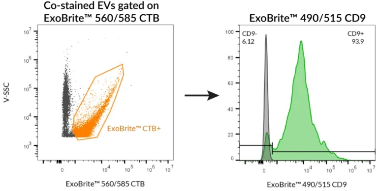
ExoBrite™ CD9 Flow Antibody is a monoclonal IgG1, kappa antibody validated by Biotium for the optimal detection of CD9, a key extracellular vesicle (EV) marker, by flow cytometry. Designed for use with both purified and bead-bound EVs, this antibody ensures bright signal, low background, and exceptional signal-to-noise performance.

Now available with an updated buffer formulation for improved staining specificity, ExoBrite™ CD9 Flow Antibody is supplied in six colour options (Pacific Blue™, FITC, PE, APC, R-PE, and APC conjugates) to support flexible multi-parameter EV analysis.

CD9, along with CD63 and CD81, belongs to the tetraspanin family, which is broadly expressed across many cell types. Its widespread presence on EVs makes CD9 a reliable biomarker for EV detection and characterisation.

Key Features

  • Optimal detection of EV marker CD9 by flow cytometry
  • Bright fluorescence, low background
  • Validated for purified and bead-bound EVs
  • Six colour options for flexible panel design
  • Updated buffer formulation for improved signal-to-noise

Notes: Biotium products are available only in Singapore and Thailand.


Detect EVs with confidence using ExoBrite™ CD9 Flow Antibody – validated for purified and bead-bound vesicles.

*Please leave us a message during checkout to indicate which kit you need, and our team will process your order accordingly.

Product Attributes

Attribute Details
Antibody number P003
SwissProt P21926
Antibody type Primary
Clonality Monoclonal
Host species Mouse
Isotype IgG1, kappa
Antibody reactivity (target) CD9
Synonyms Tetraspanin-29 (TSPAN29); BA-2/p24 antigen; BA2; BTCC1; CD9; Cell growth-inhibiting gene 2 protein; DRAP27; GIG2; Leukocyte antigen MIC3; MIC3; Motility-related protein (MRP1); p24
Species reactivity Baboon, Bovine, Cynomolgus monkey, Dog, Horse, Human, Non-human primates, Rabbit, Sheep
Human gene symbol CD9
Entrez gene ID 928
Unigene 114286
Molecular weight 24 kDa
Target cellular localisation Exosomes/EVs, Plasma membrane
Cell/tissue expression Exosomes, Platelets, Basophils, Eosinophils, Epithelial cells, Lymphocytes
Verified applications Exosome staining (verified)
Positive control MCF-7 cells, MCF-7 derived exosomes
Recommended concentration 5 µL per 0.1 mL exosomes (flow cytometry)
Research areas Exosomes/EVs
Conjugate formulation Proprietary buffer containing 0.05% sodium azide
Shelf life ≥ 24 months from receipt (if stored as recommended)
Storage conditions Store at 2–8 °C, protect fluorescent conjugates from light
Regulatory status Research Use Only (RUO)
Product origin May contain BSA (from Bos taurus serum) or recombinant BSA (CHO cells). Inquire for lot-specific details.


Variations


Antibody Ex/Em (nm) Target Species Reactivity Detection Channel Catalog No.
ExoBrite™ 410/450 CD9 Flow Antibody 416/452 CD9 Human Pacific Blue™ P003-410
ExoBrite™ 490/515 CD9 Flow Antibody 490/516 CD9 Human FITC P003-490
ExoBrite™ 560/585 CD9 Flow Antibody 562/584 CD9 Human PE P003-560
ExoBrite™ 640/660 CD9 Flow Antibody 642/663 CD9 Human APC P003-640
ExoBrite™ R-PE CD9 Flow Antibody 496/546, 565/578 CD9 Human PE P003-RPE
ExoBrite™ APC CD9 Flow Antibody 651/660 CD9 Human APC P003-APC

Available Sizes

  • 25 tests (catalogue no. suffix -125)
  • 100 tests (catalogue no. suffix -500)

Documentation

Proven Performance

SEC-purified MCF-7-derived EVs were stained with ExoBrite™ 640/660 CD9 Flow Antibody (left). Specific staining was seen, compared with the same antibody in buffer (right). Exosomes were detected on a CytoFLEX LX flow cytometer in the R660 (APC) channel.
SEC-purified MCF-7-derived EVs were stained first with ExoBrite™ 490/515 CD9 in 100 uL, followed by 10X ExoBrite™ 560/585 CTB EV Stain. EVs were detected on a CytoFLEX LX flow cytometer in the FITC and R-PE channels. When we gated on ExoBrite™ 560/585 CTB-positive particles, ~95% were also positive for CD9.
SEC-purified MCF-7-derived EVs were stained with ExoBrite™ 560/585 CD9 Flow Antibody (left). Specific staining was seen, compared with the same antibody in buffer (right). Exosomes were detected on a CytoFLEX LX flow cytometer in the Y585 (PE) channel.
SEC-purified MCF-7-derived EVs were stained with ExoBrite™ 490/515 CD9 Flow Antibody (left). Specific staining was seen, compared with the same antibody in buffer (right). Exosomes were detected on a CytoFLEX LX flow cytometer in the B525 (FITC) channel.
SEC-purified MCF-7-derived EVs were stained with ExoBrite™ 410/450 CD9 Flow Antibody (left). Specific staining was seen, compared with the same antibody in buffer (right). Exosomes were detected on a CytoFLEX LX flow cytometer in the V450 (Pacific Blue) channel.
SEC-purified MCF-7-derived EVs were stained with ExoBrite™ R-PE CD9 Flow Antibody (left). Specific staining was seen, compared with the same antibody in buffer (right). Exosomes were detected on a CytoFLEX LX flow cytometer in the Y585 (PE) channel.
EVs were isolated from MCF-7 cell supernatant with CD9 affinity beads. The bead-bound EVs were stained with ExoBrite™ CD9 490/515 Flow Antibody (green) or ExoBrite™ 490/515 IgG1 Isotype Control Flow Antibody (gray), then analyzed by flow cytometry in the FITC channel.
SEC-purified MCF-7-derived EVs were stained with ExoBrite™ APC CD9 Flow Antibody (left). Specific staining was seen, compared with the same antibody in buffer (right). Exosomes were detected on a CytoFLEX LX flow cytometer in the R660 (APC) channel.
SEC-purified HeLa-derived EVs were stained first with ExoBrite™ 560/585 CD9 in 100 uL, followed by 1X ExoBrite™ 650/665 Annexin EV Stain. EVs were detected on a CytoFLEX LX flow cytometer in the PE and APC channels. When we gated on ExoBrite™ 650/665 Annexin-positive particles, ~50% were also positive for CD9.
SEC-purified MCF-7-derived EVs were stained with ExoBrite™ 650/665 CD9 Flow Antibody (left). Specific staining was seen, compared with the same antibody in buffer (right). Exosomes were detected on a CytoFLEX LX flow cytometer in the R660 (APC) channel.
SEC-purified MCF-7-derived EVs were stained with ExoBrite™ 640/660 CD9 Flow Antibody (left). Specific staining was seen, compared with the same antibody in buffer (right). Exosomes were detected on a CytoFLEX LX flow cytometer in the R660 (APC) channel.
SEC-purified MCF-7-derived EVs were stained first with ExoBrite™ 490/515 CD9 in 100 uL, followed by 10X ExoBrite™ 560/585 CTB EV Stain. EVs were detected on a CytoFLEX LX flow cytometer in the FITC and R-PE channels. When we gated on ExoBrite™ 560/585 CTB-positive particles, ~95% were also positive for CD9.


SEC-purified MCF-7-derived EVs were stained with ExoBrite™ 560/585 CD9 Flow Antibody (left). Specific staining was seen, compared with the same antibody in buffer (right). Exosomes were detected on a CytoFLEX LX flow cytometer in the Y585 (PE) channel.


SEC-purified MCF-7-derived EVs were stained with ExoBrite™ 490/515 CD9 Flow Antibody (left). Specific staining was seen, compared with the same antibody in buffer (right). Exosomes were detected on a CytoFLEX LX flow cytometer in the B525 (FITC) channel.


SEC-purified MCF-7-derived EVs were stained with ExoBrite™ 410/450 CD9 Flow Antibody (left). Specific staining was seen, compared with the same antibody in buffer (right). Exosomes were detected on a CytoFLEX LX flow cytometer in the V450 (Pacific Blue) channel.


SEC-purified MCF-7-derived EVs were stained with ExoBrite™ R-PE CD9 Flow Antibody (left). Specific staining was seen, compared with the same antibody in buffer (right). Exosomes were detected on a CytoFLEX LX flow cytometer in the Y585 (PE) channel.


EVs were isolated from MCF-7 cell supernatant with CD9 affinity beads. The bead-bound EVs were stained with ExoBrite™ CD9 490/515 Flow Antibody (green) or ExoBrite™ 490/515 IgG1 Isotype Control Flow Antibody (gray), then analyzed by flow cytometry in the FITC channel.


SEC-purified MCF-7-derived EVs were stained with ExoBrite™ APC CD9 Flow Antibody (left). Specific staining was seen, compared with the same antibody in buffer (right). Exosomes were detected on a CytoFLEX LX flow cytometer in the R660 (APC) channel.


SEC-purified HeLa-derived EVs were stained first with ExoBrite™ 560/585 CD9 in 100 uL, followed by 1X ExoBrite™ 650/665 Annexin EV Stain. EVs were detected on a CytoFLEX LX flow cytometer in the PE and APC channels. When we gated on ExoBrite™ 650/665 Annexin-positive particles, ~50% were also positive for CD9.


SEC-purified MCF-7-derived EVs were stained with ExoBrite™ 650/665 CD9 Flow Antibody (left). Specific staining was seen, compared with the same antibody in buffer (right). Exosomes were detected on a CytoFLEX LX flow cytometer in the R660 (APC) channel.