ExoBrite™ CD63 Western Antibody

ExoBrite™ CD63 Western Antibody

ExoBrite™ CD63 Western Antibody enables bright, low-background detection of EV marker CD63 in western blots, available in fluorescent and HRP formats.

ExoBrite™ CD63 Western Antibody cover image

Add to cart

Validated Antibody for EV Marker CD63 Detection by Fluorescent or Chemiluminescent Western Blot

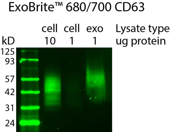
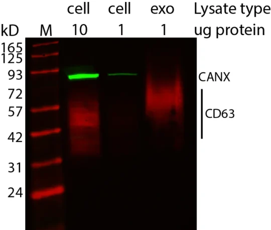
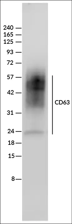
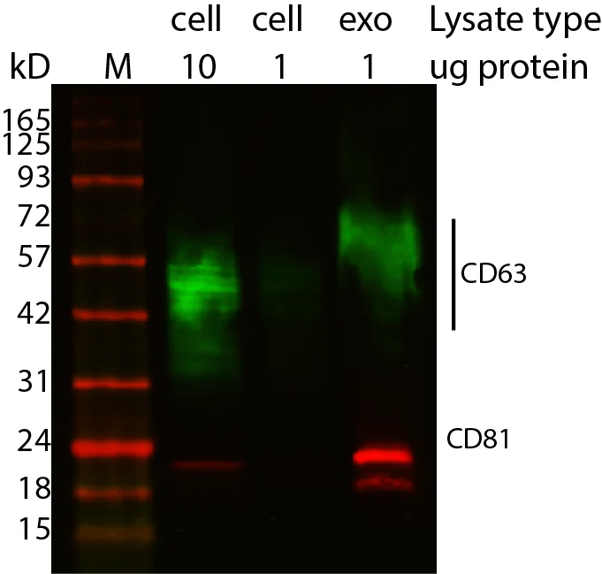
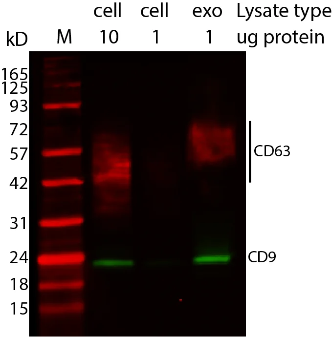
ExoBrite™ CD63 Western Antibody is a mouse monoclonal IgG1, kappa antibody validated for the detection of CD63 in extracellular vesicle (EV) extracts by western blot. Available in near-infrared fluorescent formats (680/700 and 770/800) and an HRP-conjugated format, it enables bright, low-background detection of CD63 for exosome and EV protein analysis.

Near-infrared fluorescent conjugates provide higher signal-to-noise ratios than visible-light fluorophores, making them ideal for multiplex western blotting. The HRP-conjugated format supports chemiluminescent detection for traditional workflows.

By targeting CD63, a core EV tetraspanin marker, this antibody complements ExoBrite™ CD9 and CD81 Western Antibodies, offering researchers a complete toolkit for EV characterisation.

Key Features

  • Validated for CD63 detection in EV western blots
  • Available in near-infrared fluorescent (680/700, 770/800) and HRP formats
  • Low background, bright signal for reliable EV protein analysis
  • Multiplex-capable with near-infrared detection channels
  • Complements CD9 and CD81 Western Antibodies for tetraspanin profiling


Notes: Biotium products are available only in Singapore and Thailand.


Detect CD63 in EV extracts with confidence using ExoBrite™ CD63 Western Antibody – available in fluorescent and HRP formats.

*Please leave us a message during checkout to indicate which kit you need, and our team will process your order accordingly.

Product Attributes

Attribute Details
Antibody number P004
SwissProt P08962
Antibody type Primary
Clonality Monoclonal
Host species Mouse
Isotype IgG1, kappa
Antibody reactivity (target) CD63
Synonyms gp55, granulophysin, LAMP-3, MLA1, ME491, OMA81H, PTLGP40, TSPAN30
Species reactivity Human, Baboon, Cynomolgus monkey, Non-human primates
Human gene symbol CD63
Entrez gene ID 967
Unigene 445570
Molecular weight 26 kDa (core protein); 30–60 kDa (glycosylated)
Target cellular localisation Exosomes/EVs, Lysosomes, Plasma membrane, Multivesicular bodies
Cell/tissue expression Exosomes, Platelets, Granulocytes, Lymphocytes, Monocytes/Macrophages
Verified applications Western blot (fluorescent or chemiluminescent, verified)
Positive control MCF-7 cells, MCF-7 derived exosomes
Recommended use ~100 ng/mL (1:1000 dilution for WB, adjust as required by end-user)
Conjugate formulation Fluorescent conjugates: PBS, 0.1% BSA, 0.05% azide; HRP conjugates: PBS, 50% glycerol, 2 mg/mL rBSA
Volume per assay 10 µL/test
Shelf life ≥ 24 months from receipt (if stored as recommended)
Storage conditions Store at 2–8 °C, protect fluorescent conjugates from light
Shipping condition Room temperature
Regulatory status Research Use Only (RUO)
Product origin May contain BSA (bovine) or recombinant BSA (CHO cells). Inquire for lot-specific details.

Variations

Antibody Ex/Em (nm) Concentration Size Options Detection Channel Catalog No.
ExoBrite™ 680/700 CD63 Western Antibody 681/698 100 µg/mL 25 / 100 tests NIR (700 channel) P004-680-250 / P004-680-1000
ExoBrite™ 770/800 CD63 Western Antibody 770/797 100 µg/mL 25 / 100 tests NIR (800 channel) P004-770-250 / P004-770-1000

Documentation

Proven Performance

Chemiluminescent western detection of CD63 in 10 ug of MCF7 whole cell lysate (non-reduced) using 100 ng/mL ExoBrite™ CD63-HRP conjugate, detected with SuperSignal™ West Femto Substrate (Thermo Fisher) and imaged on a LICORbio™ Odyssey® M imaging system.
Western detection of human CD63 in cell and exosome lysate using ExoBrite™ 680/700 CD63 Western Antibody, showing enrichment of CD63 in the exosome prep. Exosomes were isolated from MCF-7 cell conditioned medium by size exclusion column. The indicated amounts of lysates from MCF-7 cells and MCF-7-derived exosomes were run on an acrylamide gel and transferred to PVDF. The membrane was blocked with TrueBlack® Western Blocking Buffer, stained with 1X ExoBrite™ 680/700 CD63 Western Antibody, and imaged on a LI-COR Odyssey® infrared imaging system in the 700 channel. The protein ladder is Peacock™ Prestained Protein Marker, 1.5 uL per lane.
Western blot showing CD63 and CD81 enrichment in exosomes using ExoBrite™ Western Antibodies in MCF-7 cell and exosome lysates. Exosomes were isolated from MCF-7 cell conditioned medium by size exclusion column. The indicated amounts of lysates from MCF-7 cells and MCF-7-derived exosomes were run on an acrylamide gel and transferred to PVDF. The membrane was blocked with TrueBlack® Western Blocking Buffer, stained with 1X ExoBrite™ 680/700 CD81 Western Antibody and 1X ExoBrite™ 770/800 CD63 Western Antibody, and imaged on a LI-COR Odyssey® infrared imaging system in the 700 and 800 channels. The protein ladder is Peacock™ Plus Prestained Protein Marker, 1.5 uL loaded.
Western blot showing CD63 enrichment in exosomes using ExoBrite™ Western Antibodies in MCF-7 cell and exosome lysates. Exosomes were isolated from MCF-7 cell conditioned medium by size exclusion column. The indicated amounts of lysates from MCF-7 cells and MCF-7-derived exosomes were run on an acrylamide gel and transferred to PVDF. The membrane was blocked with TrueBlack® Western Blocking Buffer, stained with 1X ExoBrite™ 680/700 CD63 Western Antibody and 1X ExoBrite™ 770/800 Calnexin Western Antibody, and imaged on a LI-COR Odyssey® infrared imaging system in the 700 and 800 channels. The protein ladder is Peacock™ Prestained Protein Marker, 1.5 uL loaded.
Western blot showing CD9 and CD63 enrichment in exosomes using ExoBrite™ Western Antibodies in MCF-7 cell and exosome lysates. Exosomes were isolated from MCF-7 cell conditioned medium by size exclusion column. The indicated amounts of lysates from MCF-7 cells and MCF-7-derived exosomes were run on an acrylamide gel and transferred to PVDF. The membrane was blocked with TrueBlack® Western Blocking Buffer, stained with 1X ExoBrite™ 680/700 CD63 Western Antibody and 1X ExoBrite™ 770/800 CD9 Western Antibody, and imaged on a LI-COR Odyssey® infrared imaging system in the 700 and 800 channels. The protein ladder is Peacock™ Plus Prestained Protein Marker, 3 uL loaded.