ExoBrite™ CD81 Flow Antibody

ExoBrite™ CD81 Flow Antibody

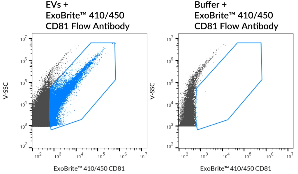
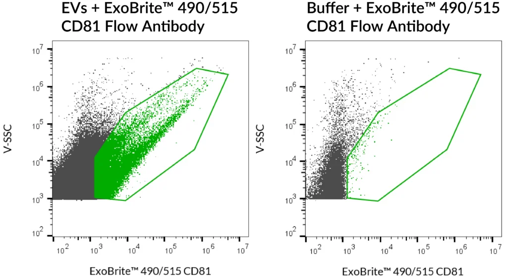
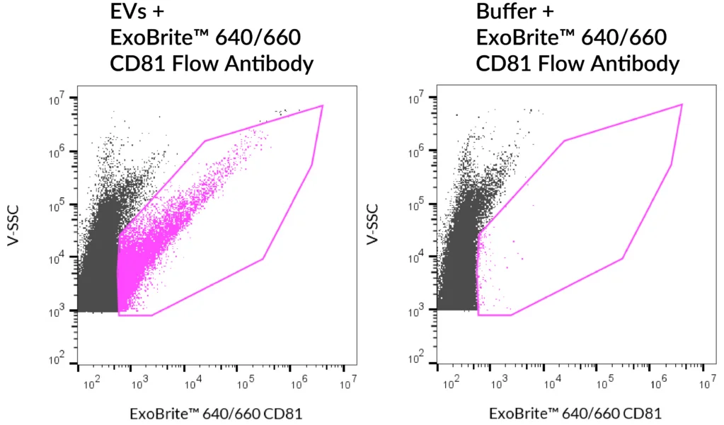
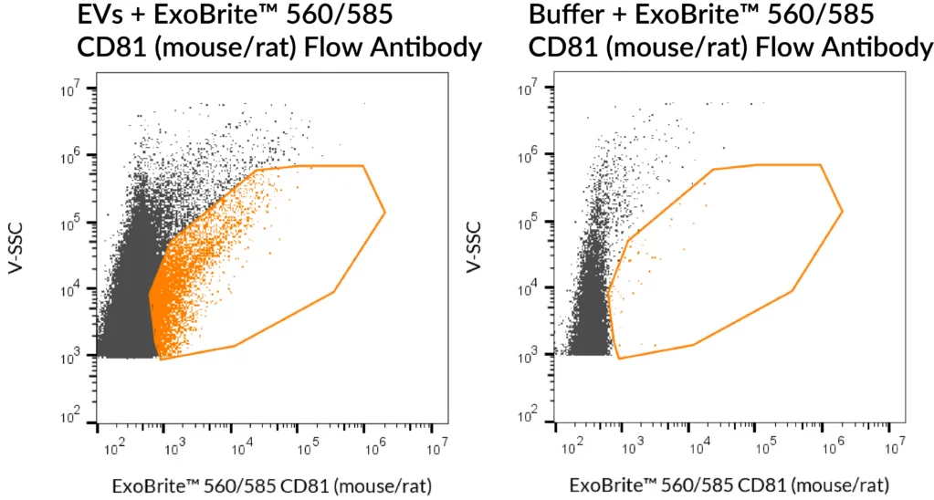
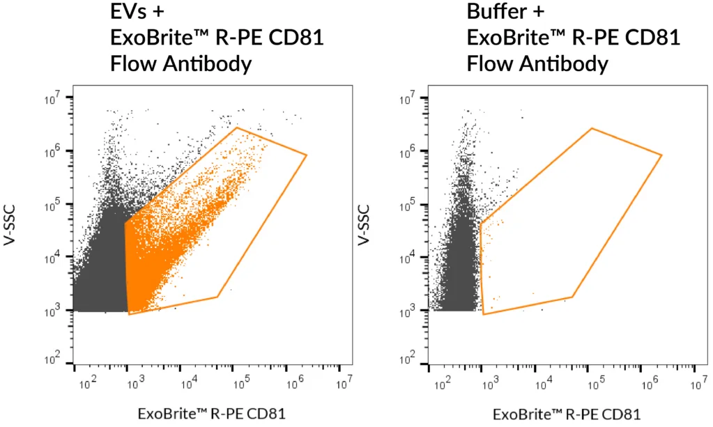
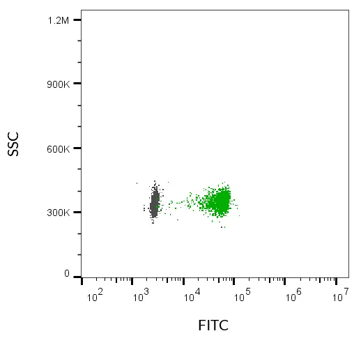
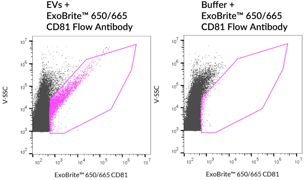
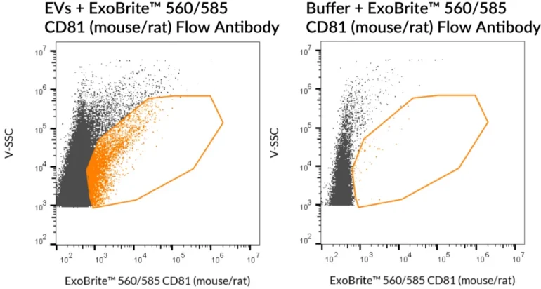
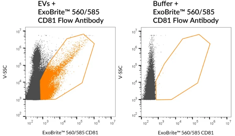
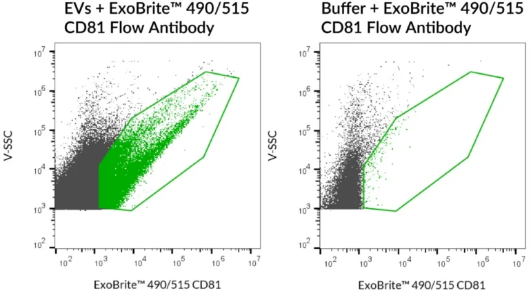
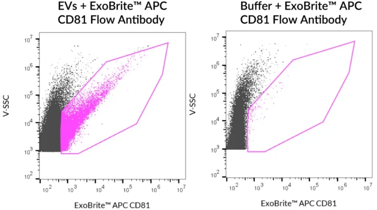
ExoBrite™ CD81 Flow Antibody provides bright, low-background detection of EV marker CD81 in purified or bead-bound EVs by flow cytometry.

ExoBrite™ CD81 Flow Antibody cover image

Add to cart

Validated Antibody for Reliable Detection of EV Marker CD81

ExoBrite™ CD81 Flow Antibody is a mouse monoclonal IgG1, kappa antibody validated by Biotium for optimal detection of CD81, a core extracellular vesicle (EV) tetraspanin marker, by flow cytometry.

Optimised for both purified and bead-bound EVs, this antibody provides bright fluorescence, low background, and superior signal-to-noise. With an updated buffer formulation, ExoBrite™ CD81 Flow Antibody ensures improved staining reproducibility and is a trusted choice for EV analysis.

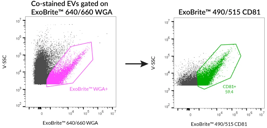
Available in six colour options (Pacific Blue™, FITC, PE, APC, R-PE, and APC conjugates), it supports multi-parameter analysis and is widely used in combination with other EV markers such as CD9 and CD63.

Key Features

  • Specific detection of EV marker CD81 by flow cytometry
  • Bright fluorescence, low background
  • Validated for purified and bead-bound EVs
  • Six colour options for panel flexibility
  • Updated buffer formulation for enhanced signal-to-noise

Notes: Biotium products are available only in Singapore and Thailand.


Detect EVs with confidence using ExoBrite™ CD81 Flow Antibody – validated for purified and bead-bound vesicles.

*Please leave us a message during checkout to indicate which kit you need, and our team will process your order accordingly.

Product Attributes

Attribute Details
Antibody number P005
SwissProt P60033
Antibody type Primary
Clonality Monoclonal
Host species Mouse
Isotype IgG1, kappa
Antibody reactivity (target) CD81
Synonyms CD81; TAPA-1; Tetraspanin-28; Tspan-28
Species reactivity Human, Baboon, Cynomolgus monkey, Non-human primates; Mouse (low reactivity)
Human gene symbol CD81
Entrez gene ID 975
Unigene 54457
Molecular weight 26 kDa
Target cellular localisation Exosomes/EVs, Plasma membrane
Cell/tissue expression Exosomes, Hepatocytes, Lymphocytes, Monocytes/Macrophages
Verified applications Exosome staining (verified)
Positive control MCF-7 cells, MCF-7 derived exosomes
Recommended concentration 5 µL per 0.1 mL exosomes (flow cytometry)
Research areas Exosomes/EVs
Conjugate formulation Proprietary buffer containing 0.05% sodium azide
Shelf life ≥ 24 months from receipt (if stored as recommended)
Storage conditions Store at 2–8 °C, protect fluorescent conjugates from light
Shipping condition Room temperature
Regulatory status Research Use Only (RUO)
Product origin May contain BSA (from bovine serum) or recombinant BSA (CHO cells). Inquire for lot-specific details.

Variations

Antibody Ex/Em (nm) Target Species Reactivity Detection Channel Catalog No.
ExoBrite™ 410/450 CD81 Flow Antibody 416/452 CD81 Human Pacific Blue™ P005-410
ExoBrite™ 490/515 CD81 Flow Antibody 490/516 CD81 Human FITC P005-490
ExoBrite™ 560/585 CD81 Flow Antibody 562/584 CD81 Human PE P005-560
ExoBrite™ 640/660 CD81 Flow Antibody 642/663 CD81 Human APC P005-640
ExoBrite™ R-PE CD81 Flow Antibody 496/546, 565/578 CD81 Human PE P005-RPE
ExoBrite™ APC CD81 Flow Antibody 651/660 CD81 Human APC P005-APC


Available Sizes

  • 25 tests (catalogue no. suffix -125)
  • 100 tests (catalogue no. suffix -500)

Documentation

Proven Performance

SEC-purified mouse NIH 3T3-derived EVs were stained with ExoBrite™ 560/585 CD81 (mouse/rat) Flow Antibody (left). Specific staining was seen, compared with the same antibody in buffer (right). Exosomes were detected on a CytoFLEX LX flow cytometer in the Y585 (PE) channel.
SEC-purified Jurkat-derived EVs were stained with ExoBrite™ 560/585 CD81 Flow Antibody (left). Specific staining was seen, compared with the same antibody in buffer (right). Exosomes were detected on a CytoFLEX LX flow cytometer in the Y585 (PE) channel.
SEC-purified MCF-7-derived EVs were stained with ExoBrite™ 490/515 CD81 Flow Antibody (left). Specific staining was seen, compared with the same antibody in buffer (right). Exosomes were detected on a CytoFLEX LX flow cytometer in the B525 (FITC) channel.
SEC-purified HEK 293-derived EVs were stained with ExoBrite™ 410/450 CD81 Flow Antibody (left). Specific staining was seen, compared with the same antibody in buffer (right). Exosomes were detected on a CytoFLEX LX flow cytometer in the V450 (Pacific Blue) channel.
SEC-purified MCF-7-derived EVs were stained with ExoBrite™ R-PE CD81 Flow Antibody (left). Specific staining was seen, compared with the same antibody in buffer (right). Exosomes were detected on a CytoFLEX LX flow cytometer in the Y585 (PE) channel.
SEC-purified MCF-7-derived EVs were stained with ExoBrite™ 490/515 CD81 Flow Antibody (left). Specific staining was seen, compared with the same antibody in buffer (right). Exosomes were detected on a CytoFLEX LX flow cytometer in the B525 (FITC) channel.
SEC-purified MCF-7-derived EVs were stained with ExoBrite™ APC CD81 Flow Antibody (left). Specific staining was seen, compared with the same antibody in buffer (right). Exosomes were detected on a CytoFLEX LX flow cytometer in the R660 (APC) channel.
SEC-purified MCF-7-derived EVs were stained with ExoBrite™ 650/665 CD81 Flow Antibody (left). Specific staining was seen, compared with the same antibody in buffer (right). Exosomes were detected on a CytoFLEX LX flow cytometer in the R660 (APC) channel.
SEC-purified MCF-7-derived EVs were stained first with ExoBrite™ 490/515 CD81 in 100 uL, followed by 1X ExoBrite™ 640/660 WGA EV Stain. EVs were detected on a CytoFLEX LX flow cytometer in the FITC and APC channels. When we gated on ExoBrite™ 640/660 WGA-positive particles, ~60% were also positive for CD81.
SEC-purified MCF-7-derived EVs were stained with ExoBrite™ 640/660 CD81 Flow Antibody (left). Specific staining was seen, compared with the same antibody in buffer (right). Exosomes were detected on a CytoFLEX LX flow cytometer in the R660 (APC) channel.