ExoBrite™ Calnexin Western Antibody

ExoBrite™ Calnexin Western Antibody

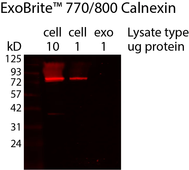
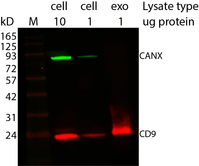
ExoBrite™ Calnexin Western Antibody validates EV purity by detecting ER protein calnexin in western blots, serving as a negative control.

ExoBrite™ Calnexin Western Antibody

Add to cart

Negative Control Antibody for Assessing EV Purity by Fluorescent Western Blot

ExoBrite™ Calnexin Western Antibody is a mouse monoclonal IgG1, kappa antibody validated for the detection of calnexin, an endoplasmic reticulum (ER) protein that is not found in extracellular vesicles (EVs).

By serving as a negative control, this antibody allows researchers to assess the purity of isolated EV extracts in western blot experiments. Offered as a conjugate to ExoBrite™ 770/800 near-infrared dye, it provides bright signal, low background, and superior signal-to-noise compared to visible-light fluorophores.

This antibody complements ExoBrite™ tetraspanin antibodies for CD9, CD63, and CD81, ensuring that EV preparations can be both positively identified and assessed for contaminating cellular proteins.

Key Features

  • Validated for detection of ER protein calnexin by fluorescent western blot
  • Serves as a negative control to confirm EV extract purity
  • Near-infrared 770/800 dye for bright, low-background detection
  • Ensures confidence in EV marker analysis by ruling out ER contamination
  • Complements CD9, CD63, and CD81 Western Antibodies for complete EV validation


Notes: Biotium products are available only in Singapore and Thailand.


Confirm the purity of your EV samples with ExoBrite™ Calnexin Western Antibody – the essential negative control for EV western blotting.

*Please leave us a message during checkout to indicate which kit you need, and our team will process your order accordingly.

Product Attributes

Attribute Details
Antibody number P007
SwissProt P27824
Antibody type Primary
Clonality Monoclonal
Host species Mouse
Isotype IgG1, kappa
Antibody reactivity (target) Calnexin
Synonyms Calnexin, CANX, CNX, IP90, p88, Major histocompatibility complex class I antigen-binding protein p88
Species reactivity Human
Human gene symbol CANX
Entrez gene ID 821
Unigene 567968
Molecular weight 67 kDa (predicted); 80–90 kDa (observed)
Target cellular localisation Endoplasmic reticulum
Cell/tissue expression All cells
Verified applications Western blot (verified)
Positive control MCF-7 cells, MCF-7 derived exosomes
Recommended use ~100 ng/mL (1:1000 dilution for WB, adjust as required by end-user)
Conjugate formulation PBS, 0.1% BSA, 0.05% azide
Volume per assay 10 µL/test
Shelf life ≥ 24 months from receipt (if stored as recommended)
Storage conditions Store at 2–8 °C, protect fluorescent conjugates from light
Shipping condition Room temperature
Regulatory status Research Use Only (RUO)
Product origin May contain BSA (bovine) or recombinant BSA (CHO cells). Inquire for lot-specific details.

Variations

Antibody Ex/Em (nm) Concentration Size Options Detection Channel Catalog No.
ExoBrite™ 770/800 Calnexin Western Antibody 770/797 100 µg/mL 25 / 100 tests NIR (800 channel) P007-770-250 / P007-770-1000

Documentation

Proven Performance

Western detection of human calnexin in cell and exosome lysate using ExoBrite™ 770/800 calnexin Western Antibody, showing the lack of calnexin in the exosome prep. Exosomes were isolated from MCF-7 cell conditioned medium by size exclusion column. The indicated amounts of lysates from MCF-7 cells and MCF-7-derived exosomes were run on an acrylamide gel and transferred to PVDF. The membrane was blocked with TrueBlack® Western Blocking Buffer, stained with 1X ExoBrite™ 770/800 calnexin Western Antibody, and imaged on a LI-COR Odyssey® infrared imaging system in the 800 cahnnel. The protein ladder is Peacock™ Prestained Protein Marker, 1.5 uL per lane.
Western blot showing CD9 enrichment in exosomes using ExoBrite™ Western Antibodies in MCF-7 cell and exosome lysates. Exosomes were isolated from MCF-7 cell conditioned medium by size exclusion column. The indicated amounts of lysates from MCF-7 cells and MCF-7-derived exosomes were run on an acrylamide gel and transferred to PVDF. The membrane was blocked with TrueBlack® Western Blocking Buffer, stained with 1X ExoBrite™ 680/700 CD9 Western Antibody and 1X ExoBrite™ 770/800 Calnexin Western Antibody, and imaged on a LI-COR Odyssey® infrared imaging system in the 700 and 800 channels. The protein ladder is Peacock™ Prestained Protein Marker, 3 uL loaded.