🔥 Don’t Miss Out on our Blind Box Giveaway with a minimum spend of $280! 🔥 Don’t Miss Out on our Blind Box Giveaway with a minimum spend of $280! 🔥 Don’t Miss Out on our Blind Box Giveaway with a minimum spend of $280!
Add friend

ExoBrite™ Annexin EV Staining Kits

ExoBrite™ Annexin EV Staining Kits

ExoBrite™ Annexin EV Staining Kits

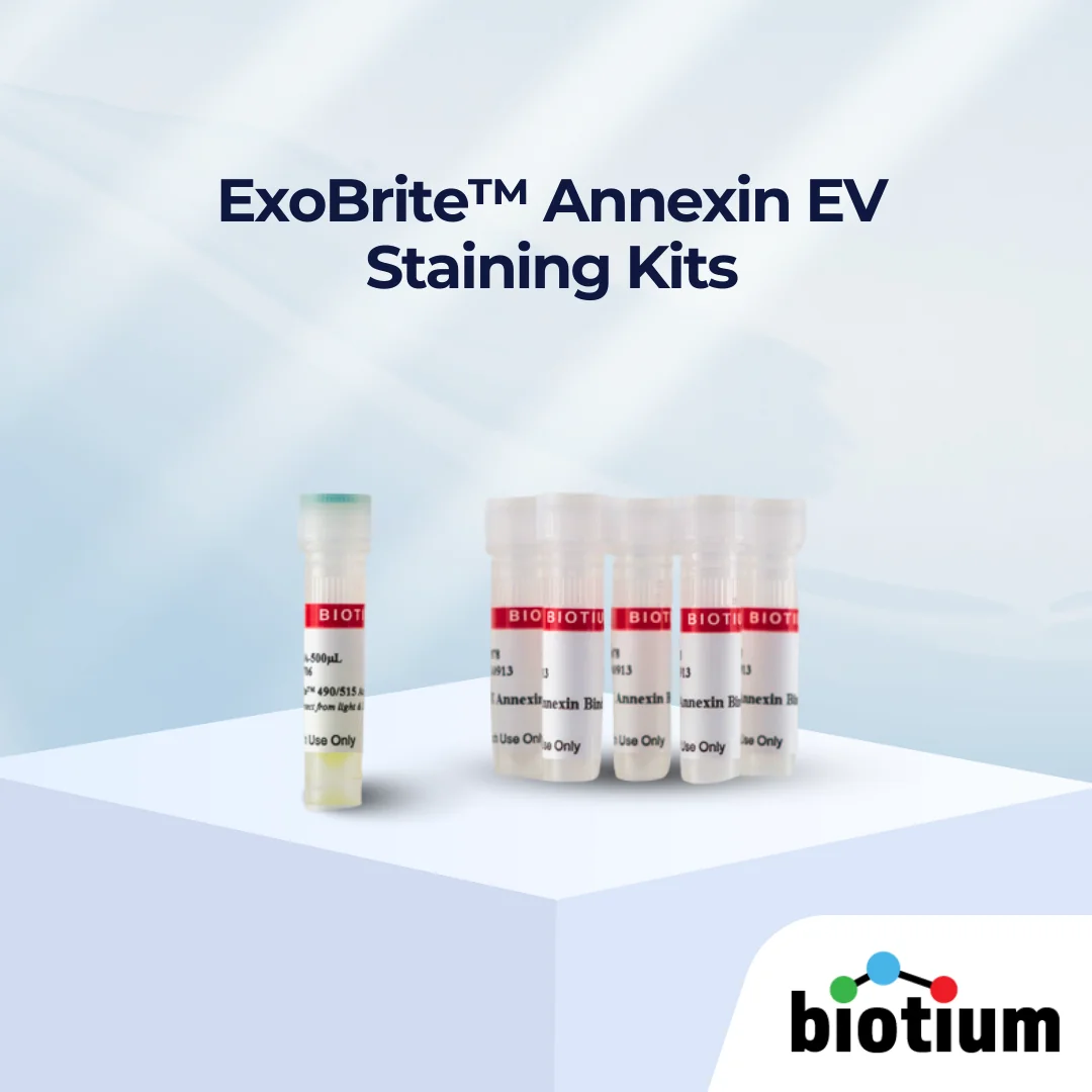
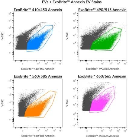
ExoBrite™ Annexin EV Staining Kits provide bright, low-background flow cytometry staining of EVs with broad compatibility across multiple sources.

ExoBrite™ Annexin EV Staining Kits cover image

Add to cart

Bright, Sensitive EV Staining with Broad Compatibility

ExoBrite™ Annexin EV Staining Kits are optimally formulated conjugates of Annexin V, designed for bright and specific staining of purified extracellular vesicles (EVs) with minimal background. Unlike traditional lipophilic dyes that risk aggregation and false signals, ExoBrite™ Annexin stains bind specifically to phosphatidylserine (PS) residues on EV membranes, enabling accurate detection by flow cytometry.

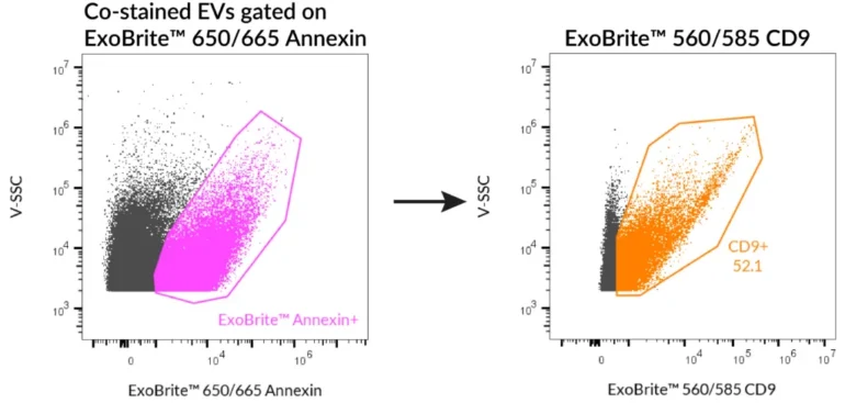
Validated across 9 different cell line-derived EV sources (MCF-7, J774, U2OS, Jurkat, HeLa, Raji, CHO, U937, A549), ExoBrite™ Annexin EV Stains provide broad coverage, strong signal intensity, and low background interference, while remaining compatible with antibody co-staining for multi-parameter EV analysis.

Each kit includes ExoBrite™ Annexin EV Stain and 50X Annexin Binding Buffer for convenient experimental setup.

Key Features

  • Optimally formulated Annexin V conjugates for EV detection
  • Bright signal, low background, designed for flow cytometry
  • Broad compatibility – stains EVs from 9 validated sources
  • Compatible with antibody co-staining (e.g. CD9, CD63, CD81)
  • Available in four colours for flexible experimental design
  • Supplied with Annexin Binding Buffer


Notes

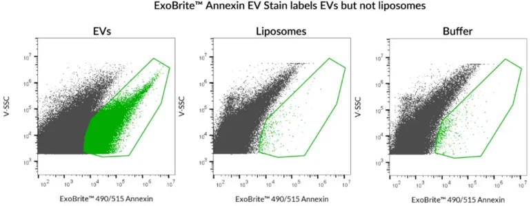
  • ExoBrite™ Annexin EV Stains label EVs from all tested cell lines, but may not stain EVs from every source.

  • For ExoBrite™ 490/515 dye, a biotin blocking step is recommended when using streptavidin-coated beads or surfaces to prevent nonspecific binding.

  • Biotium products are available only in Singapore and Thailand.


✨ Discover ExoBrite™ Annexin EV Staining Kits today – four colours are available for your EV research.


???? Please leave us a message during checkout to indicate which kit you need, and our team will process your order accordingly.

Variations

Product Ex/Em (nm) Detection Channels Size Options Catalog Number
ExoBrite™ 410/450 Annexin EV Staining Kit 416/452 nm Pacific Blue™ 100 Labelings / 500 Labelings 30119-T / 30119
ExoBrite™ 490/515 Annexin EV Staining Kit 490/516 nm FITC 100 Labelings / 500 Labelings 30120-T / 30120
ExoBrite™ 560/585 Annexin EV Staining Kit 562/584 nm PE 100 Labelings / 500 Labelings 30121-T / 30121
ExoBrite™ 650/665 Annexin EV Staining Kit 652/668 nm APC 100 Labelings / 500 Labelings 30122-T / 30122

*Please leave us a message during checkout to indicate which kit you need, and our team will process your order accordingly.

Documentation

Proven Results

SEC-purified HeLa-derived EVs were stained with ExoBrite™ Annexin EV Stains.


ExoBrite™ 490/515 Annexin EV Stain was used to stain either SEC-purified, Jurkat-derived EVs, or artificial liposomes. Robust staining was seen with the EVs, but the liposomes were not stained. The liposomes in this experiment were prepared from Presome® ACD-1, and were verified to be of similar size and concentration to the EVs using the lipophilic dye di-8-ANNEPS (data not shown). Analysis was performed on a CytoFLEX flow cytometer with SSC detected from the 405 nm laser, and ExoBrite™ detected in the FITC channel. Presome® is a registered trademark of NIPPON FINE CHEMICAL CO., LTD.
SEC-purified HeLa-derived EVs were stained first with ExoBrite™ 560/585 CD9 in 100 uL, followed by 1X ExoBrite™ 650/665 Annexin EV Stain. EVs were detected on a CytoFLEX LX flow cytometer in the PE and APC channels. When we gated on ExoBrite™ 650/665 Annexin-positive particles, ~50% were also positive for CD9.