

| Code | Description |
|---|---|
| EX05-5 | Exo-spin™, 5 mini-HD columns |
| EX05-20 | Exo-spin™, 20 mini-HD columns |
| EX05-100 | Exo-spin™, 100 mini-HD columns |
| EX06-30 | Exo-spin™ buffer, 30 ml |
| EX06-250 | Exo-spin™ buffer, 250 ml |
Product description
Exo-spin™ technology combines precipitation and size exclusion chromatography (SEC), making it superior to other techniques that rely solely on one method. Using only precipitation for exosome isolation will result in co-purification of large amounts of non-exosomal proteins and other material as well as carryover of the precipitant. SEC is reliable for exosome isolation, but a precipitation step is needed to concentrate your sample prior to SEC isolation. If there is no precipitation, a lower exosome recovery will be observed. We provide a simple two-step protocol which allows you to purify your sample with consistent and reliable results!
The EX05 Exo-spin™ kit only contains packed and equilibrated ready-to-use SEC columns, the precipitation buffer can be purchased separately (cat code EX06).
Why should I consider Exo-spin™ mini-HD?
The EX05 Exo-spin™ kit has been developed to purify exosomes from intermediate volumes, complementing the range of exosome isolation solutions.
Which Exo-spin™ kit shall I choose for my sample volume?
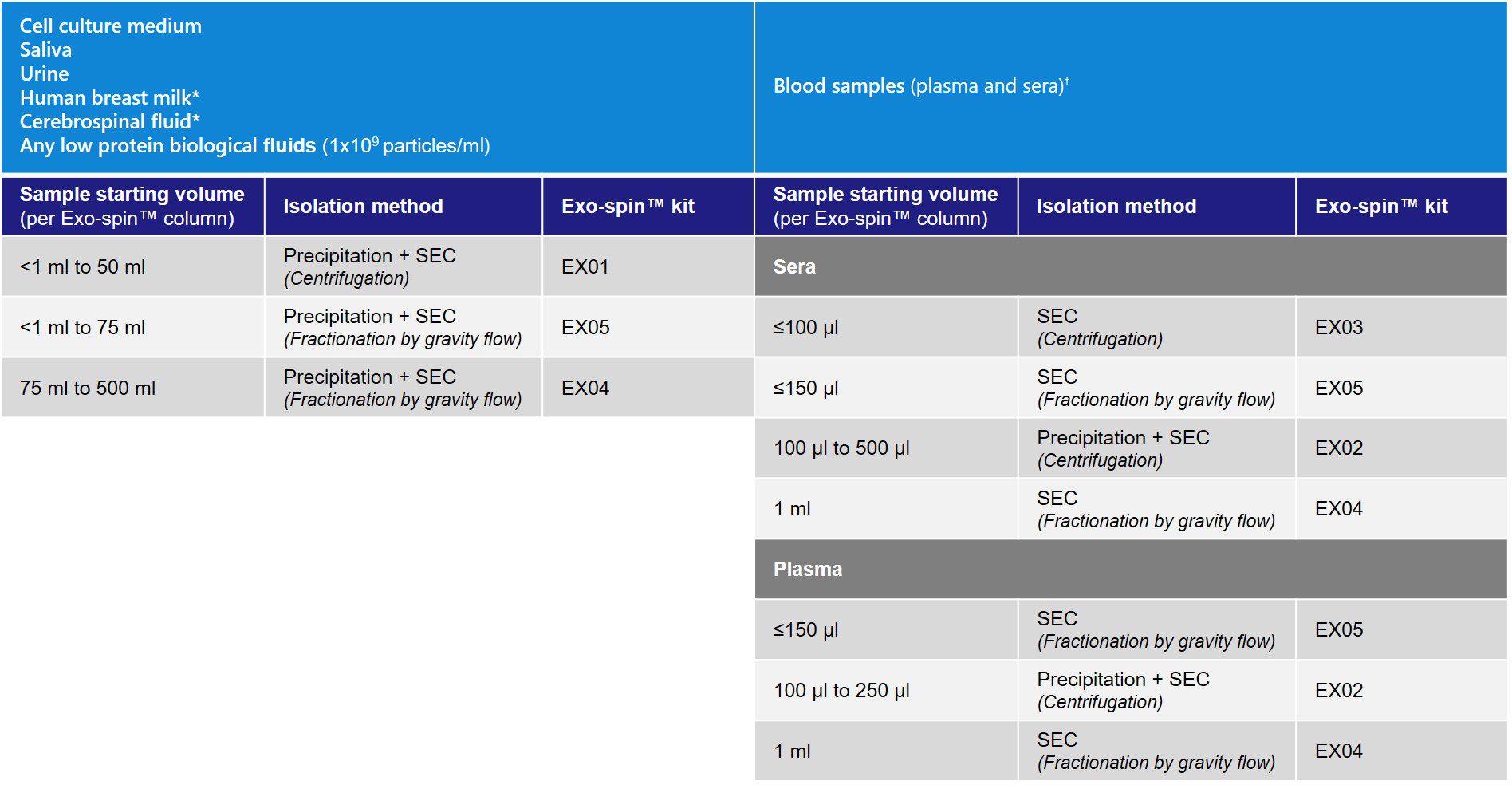
* For cerebrospinal fluid (CSF) and human breast milk samples, validated protocols are available for EX01 only. The protocols are provided in the user guides.
Highly concentrated exosome samples (e.g. 1×10¹² particles/ml) other than blood can also be used.
What is size exclusion chromatography (SEC)?
SEC is a method to separate particles in solution based on their size. The chromatography column is packed with stable polymeric beads, creating a porous matrix. When the solution containing exosomes is added to the SEC column, the smaller particles will be trapped in the pores, while larger molecules do not enter the pores and elute first. As a result, different elution fractions will contain molecules of different sizes; first the large particles, followed by the smaller particles.
This technique is ideal to achieve highly purified samples in which exosomes are separated from other non-EV components, and it is compatible with both low and high initial sample volumes.
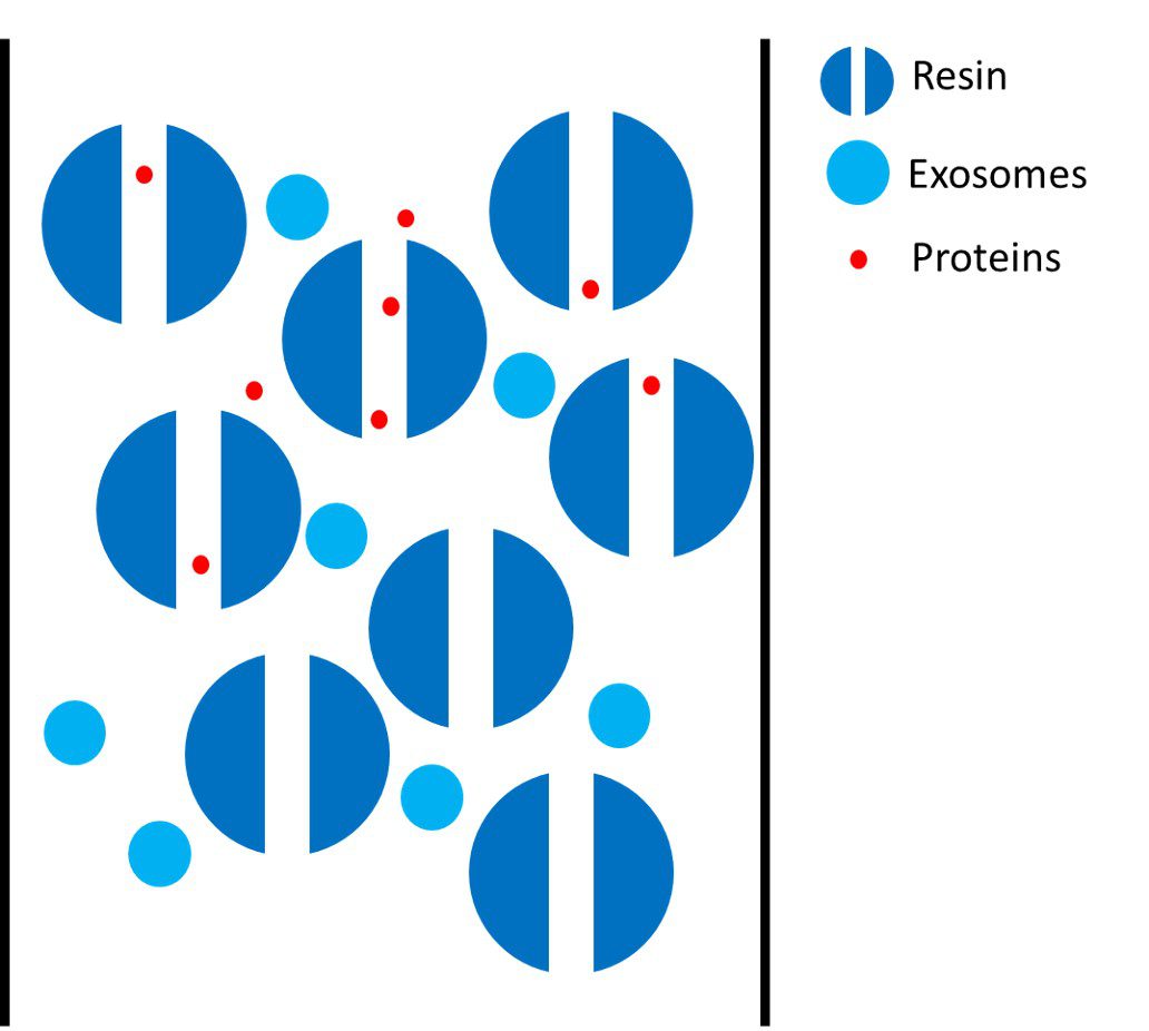
Graphic representation of the isolation of exosomes with the SEC method.
Exo-spin™ column and resin pore size
The column bed volume is 3.5 ml, allowing for 150 µl of volume to be loaded on top of the column.
The pore size of the resin is approximatively 30 nm to attain a highly pure exosome elution. All other molecules (e.g. proteins, lipids) which are smaller than 30 nm will enter into the pores and remain trapped in the column.
Fractionation analysis
The Exo-spin™ mini-HD columns allow a high-resolution fractionation protocol operated by gravity, no special equipment is required.
Highest recovery and purity
As mentioned above, essentially all proteins and lipids will be retained in the column and will elute later than the exosomes, ensuring a highly pure sample ready for your downstream application.
Reproducibility
All our columns are manufactured in our laboratory to ensure a high reproducibility between each lot. As proof of reproducibility and batch-to-batch consistency, a large number of peer-reviewed scientific papers have been published describing the use of Exo-spin™.
Storage
Upon receipt, store purification columns at 4°C.
Frequently Asked Questions (FAQs)
For any additional questions, please refer to FAQs document below.
Start today! Select our starter pack
We designed the ideal starter pack to guide your exosome research. The starter pack includes the exosome purification kit of your choice, exosome validated antibodies, and NTA profiling analysis. The complete details can be found in the product page here.
EX05 Exo-spin™ mini-HD columns data
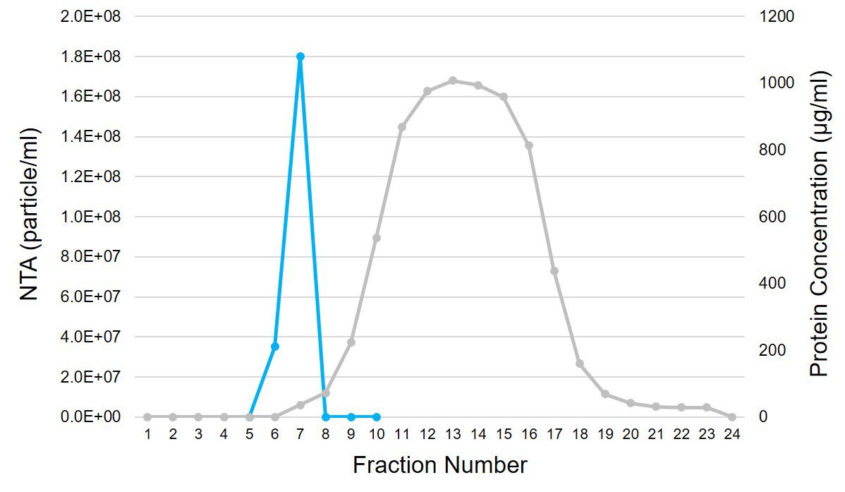
Human serum fractionation profile using Exo-spin™ mini-HD. 100 µl of human serum was added to the Exo-spin™ mini-HD column. The protein concentration was estimated by Bradford assay (grey) and the particle concentration (blue) was measured by NTA using ZetaView® instrument from Particle Metrix.
If you would like more information on the exosome extraction profile using Exo-spin™ mini-HD columns, please refer to the EX05 user guide below in the documents section.
Characterizing Exo-spin™ isolated exosomes using nanoparticle tracking analysis (NTA)
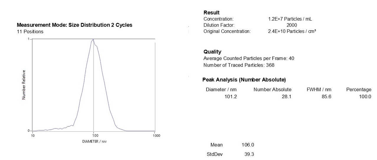
Exosome characterization with NTA. Exosomes have been isolated using the Exo-spin™ kit and analysis performed with the ZetaView® instrument.
Downstream applications: advice and content
The Exo-spin™ kits are compatible with all downstream applications and have been cited in a large range of different applications.
RNA analysis:
Immunoassay:
Exosome engineering for therapeutic cargo:
Functional study:
Proteomic (MS/MS analysis):
Protein assay:
[accordions]
[accordion title=”User guide”]EX05 Exo-spin™ user guide.pdf[/accordion]
[accordion title=”MSDS”]EX03, EX04 and EX05 MSDS.pdf [/accordion]
[accordion title=”Brochure”]CellGS exosome brochure.pdf [/accordion]
[accordion title=”FAQs”]Frequently Asked Questions (FAQs).pdf [/accordion]
Contact our Customer Care, Sales & Scientific Assistance
Consult and asked questions about our products & services
Documentation of Technical & Safety Data Sheet, Guides and more..
We gladly support you by keeping you updated on our latest products and the developments around our services.
362 Upper Paya Lebar Rd, #07-15,
Singapore 534963
WeChat ID: +65 8608 0974
© 2015-2023 Atlantis Bioscience Pte Ltd. All rights reserved. Co Reg No: 201539516N