


$357.00
Exo-spin™ technology combines precipitation and size exclusion chromatography (SEC), making it superior to other techniques that rely solely on one method. Using only precipitation for exosome isolation will result in co-purification of large amounts of non-exosomal proteins and other material as well as carryover of the precipitant. SEC is reliable for exosome isolation, but a precipitation step is needed to concentrate your sample prior to SEC isolation. If there is no precipitation, a lower exosome recovery will be observed. We provide a simple two-step protocol which allows you to purify your sample in less than 2 hours with consistent and reliable results!
The EX02 Exo-spin™ kit has been developed to process small volumes of blood samples (both plasma and sera). The kit includes Exo-spin™ buffer as well as SEC columns. The Exo-spin™ buffer is a polymer-based precipitation buffer and will be used for the first step. For the second step, pre-packed and equilibrated ready-to-use SEC columns are included.
| Code | Description |
|---|---|
| EX02-8 | Exo-spin™ blood, 8 columns, 2 ml buffer |
| EX02-25 | Exo-spin™ blood, 24 columns, 25 ml buffer |
| EX02-50 | Exo-spin™ blood, 24 columns, 30 ml buffer |
| EX06-30 | Exo-spin™ buffer, 30 ml |
| EX06-250 | Exo-spin™ buffer, 250 ml |
Which Exo-spin™ kit shall I choose for my sample volume?
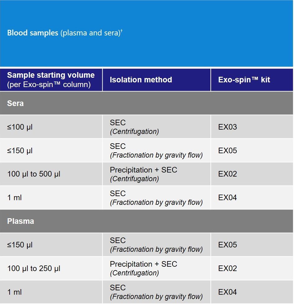
† Highly concentrated exosome samples (e.g. 1×10¹² particles/ml) other than blood can also be used.
Exo-spin™ column and resin pore size
The column bed volume is 500 µl, allowing for 100 µl of volume to be loaded on top of the column.
The pore size of the resin is approximatively 30 nm to attain a highly pure exosome elution. All other molecules (e.g. proteins, lipids) which are smaller than 30 nm will enter into the pores and remain trapped in the column.
Highest recovery and purity
As mentioned above, essentially all proteins and lipids will be retained in the column and will elute later than the exosomes, ensuring a highly pure sample ready for your downstream application.
All our columns are manufactured in our laboratory to ensure a high reproducibility between each lot. As proof of reproducibility and batch-to-batch consistency, a large number of peer-reviewed scientific papers have been published describing the use of Exo-spin™.
Upon receipt, store purification columns and Exo-spin™ Buffer at 4°C. All other components should be stored at room temperature (15°C – 25°C).
Frequently Asked Questions (FAQs)
For any additional questions, please refer to FAQs document below.
Start today! Select our starter pack
We designed the ideal starter pack to guide your exosome research. The starter pack includes the exosome purification kit of your choice, exosome validated antibodies, and NTA profiling analysis. The complete details can be found in the product page here.
[accordions]
[accordion title=”Product Data”]
EX02 Exo-spin™ kit comparative data
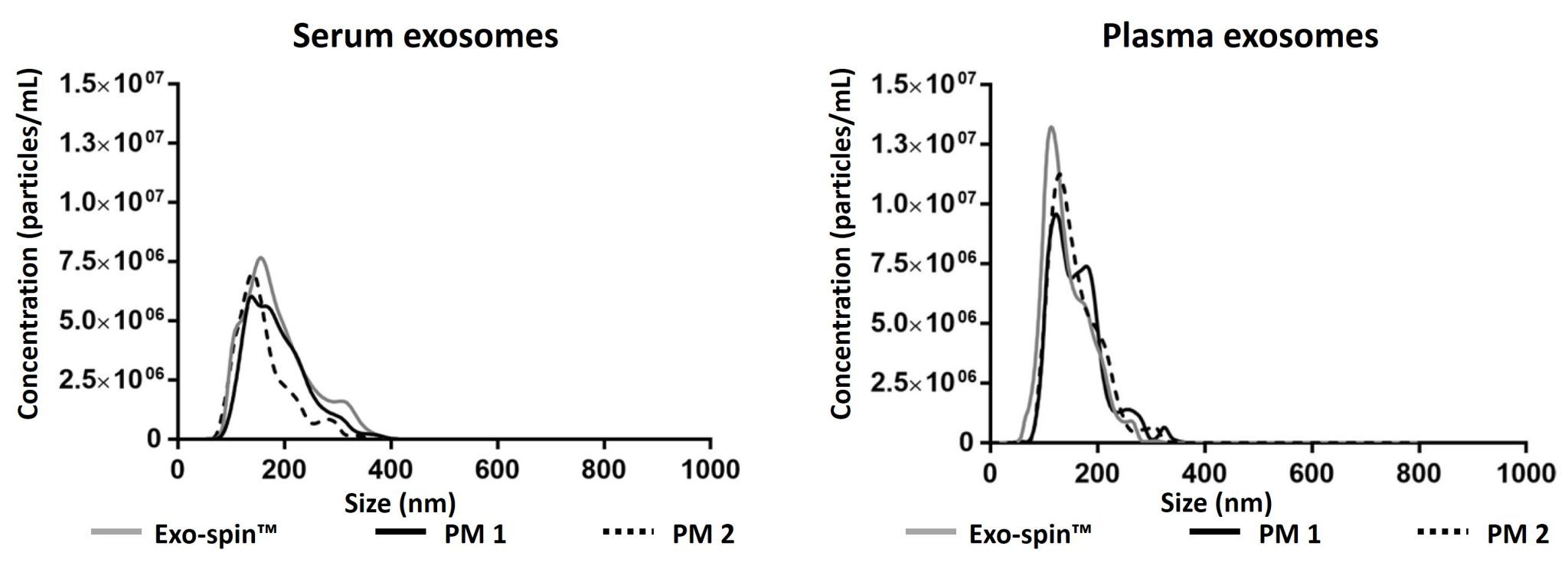
Data determined by nanoparticle tracking analysis (NTA). Each curve represents the average of 3 technical replicate measurements for each exosome isolation method and biofluid triplicate experiment. (PM = Precipitation Method). Figure taken and adapted from (Martins, TS et al., 2018).
Exosome characterization with NTA. Exosomes have been isolated using the Exo-spin™ kit and analysis performed with the ZetaView® instrument.
Characterizing Exo-spin™ isolated exosomes using nanoparticle tracking analysis (NTA)
Exosome characterization with NTA. Exosomes have been isolated using the Exo-spin™ kit and analysis performed with the ZetaView® instrument.
[/accordion]
[accordions]
[accordion title=”Downstream applications”]
The EX02 Exo-spin™ kit is compatible with all downstream application and has been published in a large range of different applications.
RNA analysis:
Mass Spectrometry:
Functional study:
Application note:
[/accordion]
[accordion title=”User guide”] EX02_Exo-spin_User_Guide[/accordion]
[accordion title=”FAQs”] FAQs[/accordion]
[accordion title=”MSDS/Tech Sheet”] EX01 and EX02 MSDS.pdf[/accordion]
[accordion title=”Product Brochure”] CellGS exosome brochure.pdf[/accordion]
[/accordions]
Contact our Customer Care, Sales & Scientific Assistance
Consult and asked questions about our products & services
Documentation of Technical & Safety Data Sheet, Guides and more..
We gladly support you by keeping you updated on our latest products and the developments around our services.
362 Upper Paya Lebar Rd, #07-15,
Singapore 534963
WeChat ID: +65 8608 0974
© 2015-2023 Atlantis Bioscience Pte Ltd. All rights reserved. Co Reg No: 201539516N