🔥 Don’t Miss Out on our Blind Box Giveaway with a minimum spend of $280! 🔥 Don’t Miss Out on our Blind Box Giveaway with a minimum spend of $280! 🔥 Don’t Miss Out on our Blind Box Giveaway with a minimum spend of $280!
Add friend

CytoLiner™ Fixed Cell Membrane Stains

CytoLiner™ Fixed Cell Membrane Stains

CytoLiner™ Fixed Cell Membrane Stains

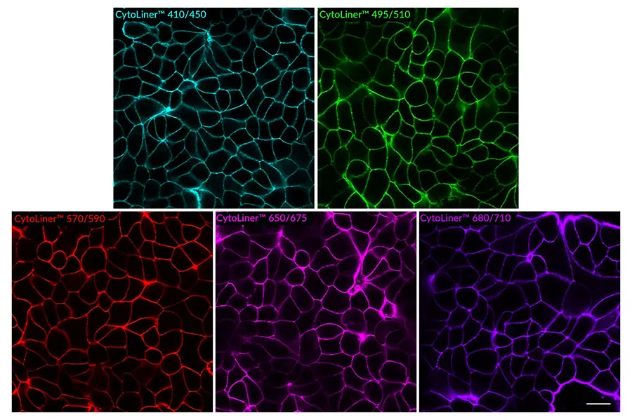
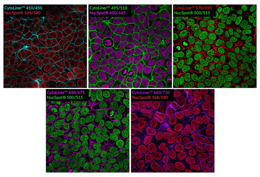
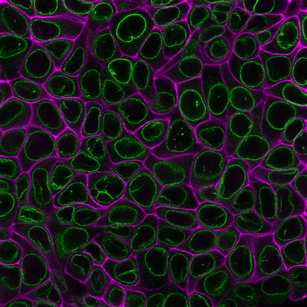
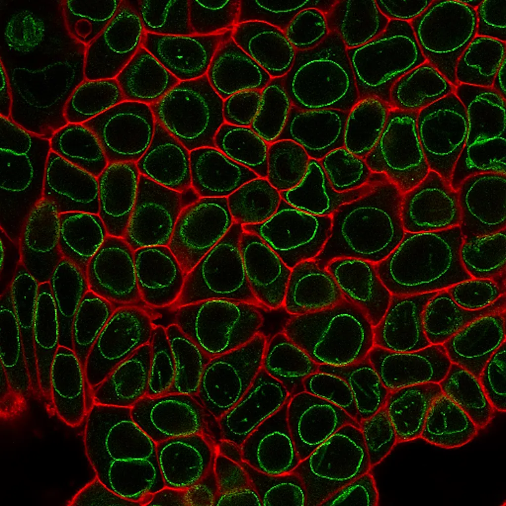
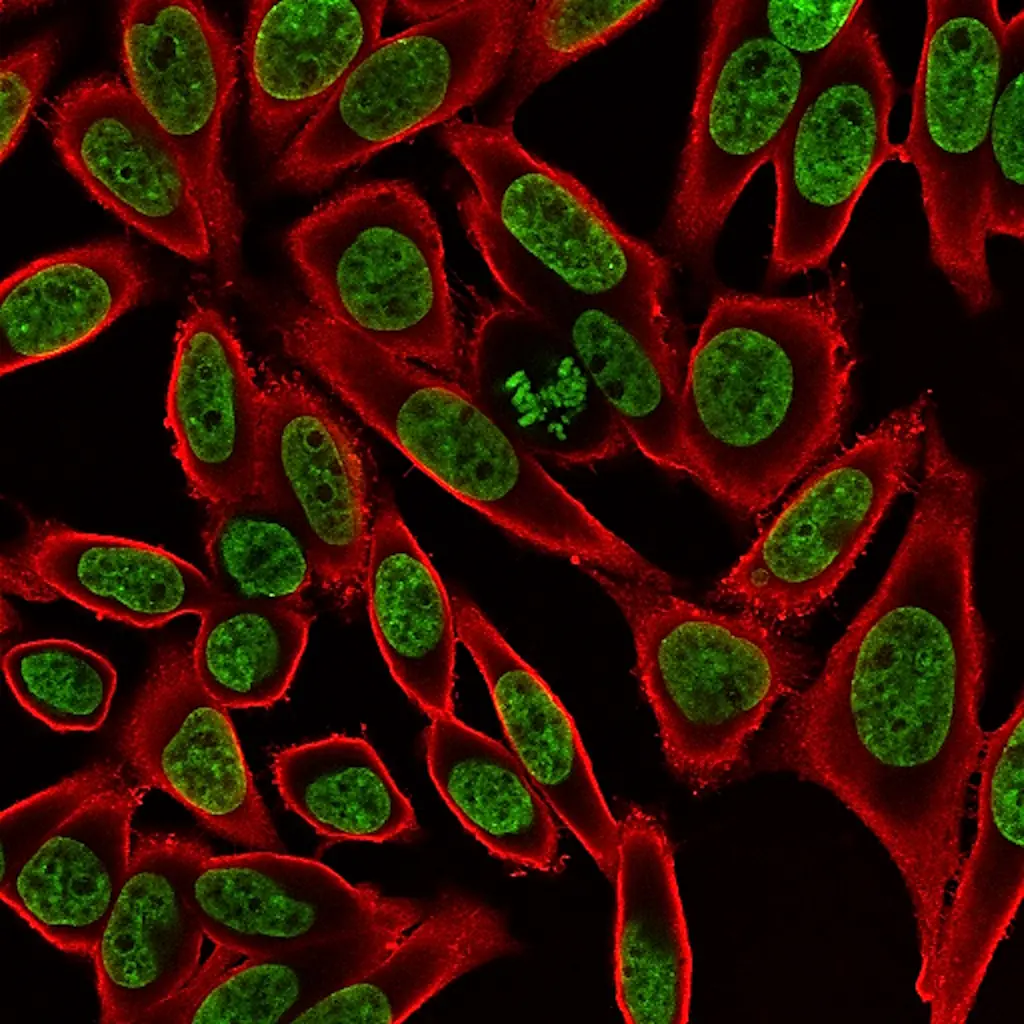
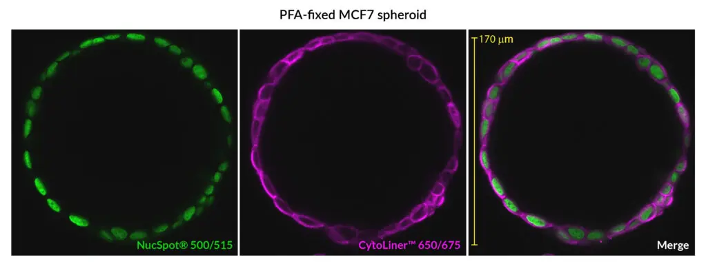
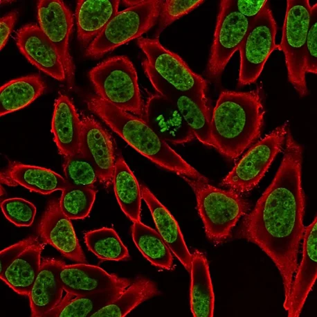
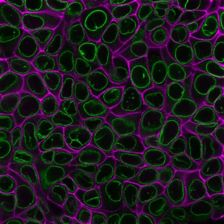
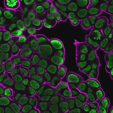
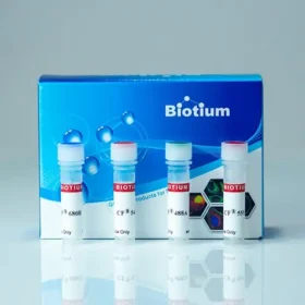
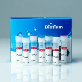
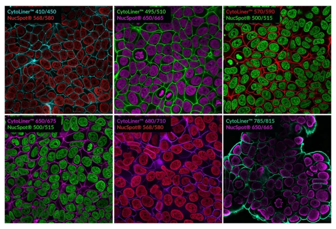
CytoLiner™ Fixed Cell Membrane Stains are novel lipophilic fluorescent dyes engineered for robust and consistent plasma membrane staining in formaldehyde-fixed cells. Available in six colours from blue to near-infrared, these dyes are optimised for microscopy and immunofluorescence workflows.

CytoLiner™ Fixed Cell Membrane Stains Cover image

Add to cart
SKU: 30131-T, 30131, 30132-T, 30132, 30133-T, 30133, 30134-T, 30134, 30135-T, 30135, 30140-T, 30140 Categories: Brand:

Novel Dyes for Reliable Membrane Labelling

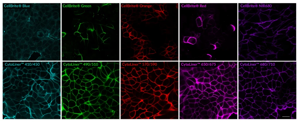
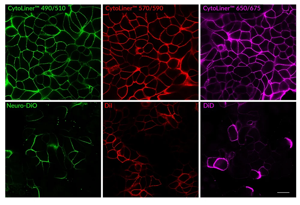
CytoLiner™ Fixed Cell Membrane Dyes represent a new generation of lipophilic fluorescent dyes specifically developed for selective plasma membrane staining in fixed and mildly permeabilised cells. Unlike traditional carbocyanine dyes such as DiI, which often show poor solubility and inconsistent staining, CytoLiner™ dyes are uniquely formulated for reliable and uniform membrane labelling in formaldehyde-fixed samples.

These dyes are compatible with immunofluorescence protocols, tolerating blocking agents, detergents, and antibody co-staining. They can also be used with poly-L-lysine coated cultureware and Transwell® supports, ensuring flexibility across a wide range of microscopy applications.

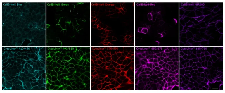
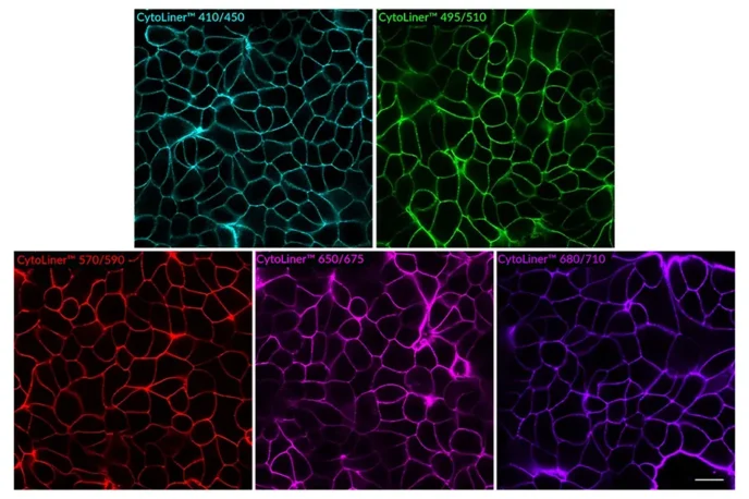
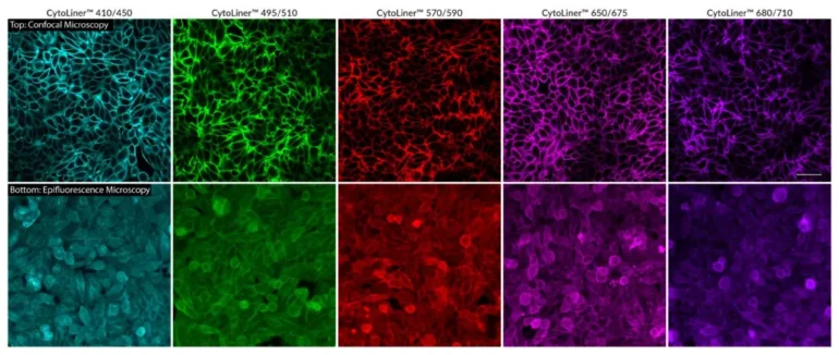
CytoLiner™ dyes are available in six excitation/emission pairs covering blue to near-IR detection channels, making them suitable for multicolour imaging panels.

Key Features

  • Robust, consistent plasma membrane staining in formaldehyde-fixed cells
  • Optimised for downstream antibody co-staining and multiplex imaging
  • Compatible with poly-L-lysine coated plates and Transwell® inserts
  • Works with mild detergent permeabilisation and blocking agents
  • Available in six spectral variants (410/450 to 785/815 nm)
  • Supplied as 500X dye in DMSO plus 100X staining buffer


Superior Staining Performance Over Classic Membrane Dyes

Comparison of fixed cell staining with CytoLiner™ Fixed Cell Membrane Stains versus classic lipophilic carbocyanine membrane dyes. In comparison to the traditional lipophilic carbocyanine dyes, CytoLiner™ dyes show more uniform and reliable staining of PFA-fixed, mildly-permeabilized cell membranes. PFA-fixed MCF7 cells were permeabilized for 10 minutes at RT with PBS/0.1% Triton® X-100, then rinsed with PBS and stained with carbocyanine dyes in PBS or CytoLiner™ Stains in 1X CytoLiner™ Buffer for 10 minutes at RT, then rinsed with PBS. Imaged by confocal microscopy; scale bar: 20 um.

Suitable for Co-Staining with Antibodies and Other Probes

PFA-fixed MCF-7 cells were permeabilized with PBS/0.1% Triton® X-100 for 10 minutes at room temperature, then stained with 1X CytoLiner™ Fixed Cell Membrane Dyes and NucSpot® Nuclear Stains.


Kit Components:

  • CytoLiner™ Fixed Cell Membrane Dye (500X in DMSO)
  • CytoLiner™ Fixed Cell Staining Buffer (100X)

Notes: Biotium products are available only in Singapore and Thailand.

*Please leave us a message during checkout to indicate which kit you need, and our team will process your order accordingly.

Variation

Variant Excitation/Emission (nm) Detection Channels Cat. No. (1000 Labelings) Cat. No. (250 Labelings)
CytoLiner™ 410/450 406/446 DAPI/Pacific Blue™ 30131 30131-T
CytoLiner™ 495/510 492/510 FITC 30132 30132-T
CytoLiner™ 570/590 573/592 Cy®3/TRITC 30133 30133-T
CytoLiner™ 650/675 647/674 Cy®5 30134 30134-T
CytoLiner™ 680/710 682/707 Cy®5.5 30135 30135-T
CytoLiner™ 785/815 787/819 Alexa Fluor® 790 30140 30140-T

Proven Performance

Staining of PFA-fixed MCF7 spheroid by NucSpot® 500/515 (green) and CytoLiner™ 650/675 (magenta). MCF-7 cells were grown in a cell repellent U-bottom 96-well plate for 17 days to allow spheroid formation, then fixed with 4% PFA. Cells were permeabilized for 10 minutes at room temperature with PBS/0.1% Triton® X-100 and stained with 1X NucSpot® and 1X CytoLiner™ in 1X CytoLiner™ Staining Buffer for 10 minutes at room temperature, washed, and imaged by confocal microscopy using an Evident FV4000 imaging system.


Staining of cells on Transwell® cell culture insert with NucSpot® 500/515 (green) and CytoLiner™ 570/590 (red). HeLa cells were grown on a 6-well Transwell® insert. The insert with cells was fixed in the microplate with 4% PFA, then permeabilized for 10 minutes at room temperature with PBS/0.1% Triton X-100 and washed with PBS. The insert was stained with 1X NucSpot® and 1X CytoLiner™ in 1X CytoLiner™ Staining Buffer for 10 minutes at room temperature and washed with PBS. The insert was cut from the support using a scalpel and mounted on a slide under a coverslip with 1% methylcellulose in PBS, then imaged by confocal microscopy.


Comparison of fixed cell staining with CytoLiner™ Fixed Cell Membrane Stains versus the classic CellBrite® Cytoplasmic Membrane Dyes. In comparison to the CellBrite® lipophilic carbocyanine dyes, CytoLiner™ dyes show more uniform and reliable staining of PFA-fixed, mildly-permeabilized cell membranes. PFA-fixed cells were permeabilized for 10 minutes at RT with PBS/0.1% Triton® X-100, then rinsed with PBS and stained with CellBrite® Dyes in PBS or CytoLiner™ Stains in 1X CytoLiner™ Buffer for 10 minutes at RT, then rinsed with PBS. Imaged by confocal microscopy; scale bar: 20 um


Comparison of fixed cell staining with CytoLiner™ Fixed Cell Membrane Stains versus classic lipophilic carbocyanine membrane dyes. In comparison to the traditional lipophilic carbocyanine dyes, CytoLiner™ dyes show more uniform and reliable staining of PFA-fixed, mildly-permeabilized cell membranes. PFA-fixed MCF7 cells were permeabilized for 10 minutes at RT with PBS/0.1% Triton® X-100, then rinsed with PBS and stained with carbocyanine dyes in PBS or CytoLiner™ Stains in 1X CytoLiner™ Buffer for 10 minutes at RT, then rinsed with PBS. Imaged by confocal microscopy; scale bar: 20 um.


PFA-fixed MCF-7 cells were permeabilized with PBS/0.1% Triton® X-100 for 10 minutes at room temperature, then stained with 1X CytoLiner™ Fixed Cell Membrane Dyes and NucSpot® Nuclear Stains.


PFA-fixed MCF-7 cells were permeabilized with PBS/0.1% Triton® X-100 for 10 minutes at room temperature, then stained with 1X CytoLiner™ Fixed Cell Membrane Dye diluted in 1X CytoLiner™ Fixed Cell Staining Buffer for 10 minutes at room temperature, followed by two rinses in PBS. Cells were imaged in PBS by confocal microscopy. Scale bar: 20 um.


PFA-fixed HeLa cells were permeabilized with PBS/0.1% Triton® X-100 for 10 minutes at room temperature, then stained with 1X CytoLiner™ Fixed Cell Membrane Dye diluted in 1X CytoLiner™ Fixed Cell Staining Buffer for 10 minutes at room temperature, followed by two rinses in PBS. Cells were imaged in PBS by confocal microscopy or epifluorescnence microscopy. Confocal microscopy permits clearer images of cell boundaries in a narrow focal plane, while epifluorescence microscopy captures membrane fluorescence at muliple focal planes of the cell. Scale bar: 50 um for confocal images.


PFA-fixed MCF7 cells were permeabilized for 10 minutes at RT with 0.1% Triton® X-100 in PBS and stained with 1X CytoLiner™ 650/675 dye in 1X CytoLiner™ Staining Buffer. Cells were then blocked with PBS/2% fish gelatin and stained with 5 ug/mL CF488A Recombinant Anti-Nuclear Membrane Antibody (clone 2406.NM) in the same buffer used for blocking. CytoLiner™ staining is shown in magenta, nuclear membrane antibody staining is shown in green.


PFA-fixed MCF7 cells were permeabilized for 10 minutes at RT with 0.1% Triton® X-100 in PBS and stained with 1X CytoLiner™ 570/590 dye in 1X CytoLiner™ Staining Buffer. Cells were then blocked with PBS/2% fish gelatin and stained with 5 ug/mL CF488A Recombinant Anti-Nuclear Membrane Antibody (clone 2406.NM) in the same buffer used for blocking. CytoLiner™ staining is shown in red, nuclear membrane antibody staining is shown in green.
Staining of cells on Transwell® cell culture insert with NucSpot® 500/515 (green) and CytoLiner™ 570/590 (red). HeLa cells were grown on a 6-well Transwell® insert. The insert with cells was fixed in the microplate with 4% PFA, then permeabilized for 10 minutes at room temperature with PBS/0.1% Triton X-100 and washed with PBS. The insert was stained with 1X NucSpot® and 1X CytoLiner™ in 1X CytoLiner™ Staining Buffer for 10 minutes at room temperature and washed with PBS. The insert was cut from the support using a scalpel and mounted on a slide under a coverslip with 1% methylcellulose in PBS, then imaged by confocal microscopy.
Comparison of fixed cell staining with CytoLiner™ Fixed Cell Membrane Stains versus the classic CellBrite® Cytoplasmic Membrane Dyes. In comparison to the CellBrite® lipophilic carbocyanine dyes, CytoLiner™ dyes show more uniform and reliable staining of PFA-fixed, mildly-permeabilized cell membranes. PFA-fixed cells were permeabilized for 10 minutes at RT with PBS/0.1% Triton® X-100, then rinsed with PBS and stained with CellBrite® Dyes in PBS or CytoLiner™ Stains in 1X CytoLiner™ Buffer for 10 minutes at RT, then rinsed with PBS. Imaged by confocal microscopy; scale bar: 20 um
Comparison of fixed cell staining with CytoLiner™ Fixed Cell Membrane Stains versus classic lipophilic carbocyanine membrane dyes. In comparison to the traditional lipophilic carbocyanine dyes, CytoLiner™ dyes show more uniform and reliable staining of PFA-fixed, mildly-permeabilized cell membranes. PFA-fixed MCF7 cells were permeabilized for 10 minutes at RT with PBS/0.1% Triton® X-100, then rinsed with PBS and stained with carbocyanine dyes in PBS or CytoLiner™ Stains in 1X CytoLiner™ Buffer for 10 minutes at RT, then rinsed with PBS. Imaged by confocal microscopy; scale bar: 20 um.
PFA-fixed MCF-7 cells were permeabilized with PBS/0.1% Triton® X-100 for 10 minutes at room temperature, then stained with 1X CytoLiner™ Fixed Cell Membrane Dyes and NucSpot® Nuclear Stains.
PFA-fixed MCF-7 cells were permeabilized with PBS/0.1% Triton® X-100 for 10 minutes at room temperature, then stained with 1X CytoLiner™ Fixed Cell Membrane Dye diluted in 1X CytoLiner™ Fixed Cell Staining Buffer for 10 minutes at room temperature, followed by two rinses in PBS. Cells were imaged in PBS by confocal microscopy. Scale bar: 20 um.
PFA-fixed HeLa cells were permeabilized with PBS/0.1% Triton® X-100 for 10 minutes at room temperature, then stained with 1X CytoLiner™ Fixed Cell Membrane Dye diluted in 1X CytoLiner™ Fixed Cell Staining Buffer for 10 minutes at room temperature, followed by two rinses in PBS. Cells were imaged in PBS by confocal microscopy or epifluorescnence microscopy. Confocal microscopy permits clearer images of cell boundaries in a narrow focal plane, while epifluorescence microscopy captures membrane fluorescence at muliple focal planes of the cell. Scale bar: 50 um for confocal images.
PFA-fixed MCF7 cells were permeabilized for 10 minutes at RT with 0.1% Triton® X-100 in PBS and stained with 1X CytoLiner™ 650/675 dye in 1X CytoLiner™ Staining Buffer. Cells were then blocked with PBS/2% fish gelatin and stained with 5 ug/mL CF488A Recombinant Anti-Nuclear Membrane Antibody (clone 2406.NM) in the same buffer used for blocking. CytoLiner™ staining is shown in magenta, nuclear membrane antibody staining is shown in green.
PFA-fixed MCF7 cells were permeabilized for 10 minutes at RT with 0.1% Triton® X-100 in PBS and stained with 1X CytoLiner™ 570/590 dye in 1X CytoLiner™ Staining Buffer. Cells were then blocked with PBS/2% fish gelatin and stained with 5 ug/mL CF488A Recombinant Anti-Nuclear Membrane Antibody (clone 2406.NM) in the same buffer used for blocking. CytoLiner™ staining is shown in red, nuclear membrane antibody staining is shown in green.
Staining of cells on Transwell® cell culture insert with NucSpot® 500/515 (green) and CytoLiner™ 650/675 (magenta). MCF7 cells were grown on a 6-well Transwell® insert. The insert with cells was fixed in the microplate with 4% PFA, then permeabilized for 10 minutes at room temperature with PBS/0.1% Triton X-100 and washed with PBS. The insert was stained with 1X NucSpot® and 1X CytoLiner™ in 1X CytoLiner™ Staining Buffer for 10 minutes at room temperature and washed with PBS. The insert was cut from the support using a scalpel and mounted on a slide under a coverslip with 1% methylcellulose in PBS, then imaged by confocal microscopy.
Staining of PFA-fixed MCF7 spheroid by NucSpot® 500/515 (green) and CytoLiner™ 650/675 (magenta). MCF-7 cells were grown in a cell repellent U-bottom 96-well plate for 17 days to allow spheroid formation, then fixed with 4% PFA. Cells were permeabilized for 10 minutes at room temperature with PBS/0.1% Triton® X-100 and stained with 1X NucSpot® and 1X CytoLiner™ in 1X CytoLiner™ Staining Buffer for 10 minutes at room temperature, washed, and imaged by confocal microscopy using an Evident FV4000 imaging system.


Staining of cells on Transwell® cell culture insert with NucSpot® 500/515 (green) and CytoLiner™ 650/675 (magenta). MCF7 cells were grown on a 6-well Transwell® insert. The insert with cells was fixed in the microplate with 4% PFA, then permeabilized for 10 minutes at room temperature with PBS/0.1% Triton X-100 and washed with PBS. The insert was stained with 1X NucSpot® and 1X CytoLiner™ in 1X CytoLiner™ Staining Buffer for 10 minutes at room temperature and washed with PBS. The insert was cut from the support using a scalpel and mounted on a slide under a coverslip with 1% methylcellulose in PBS, then imaged by confocal microscopy.