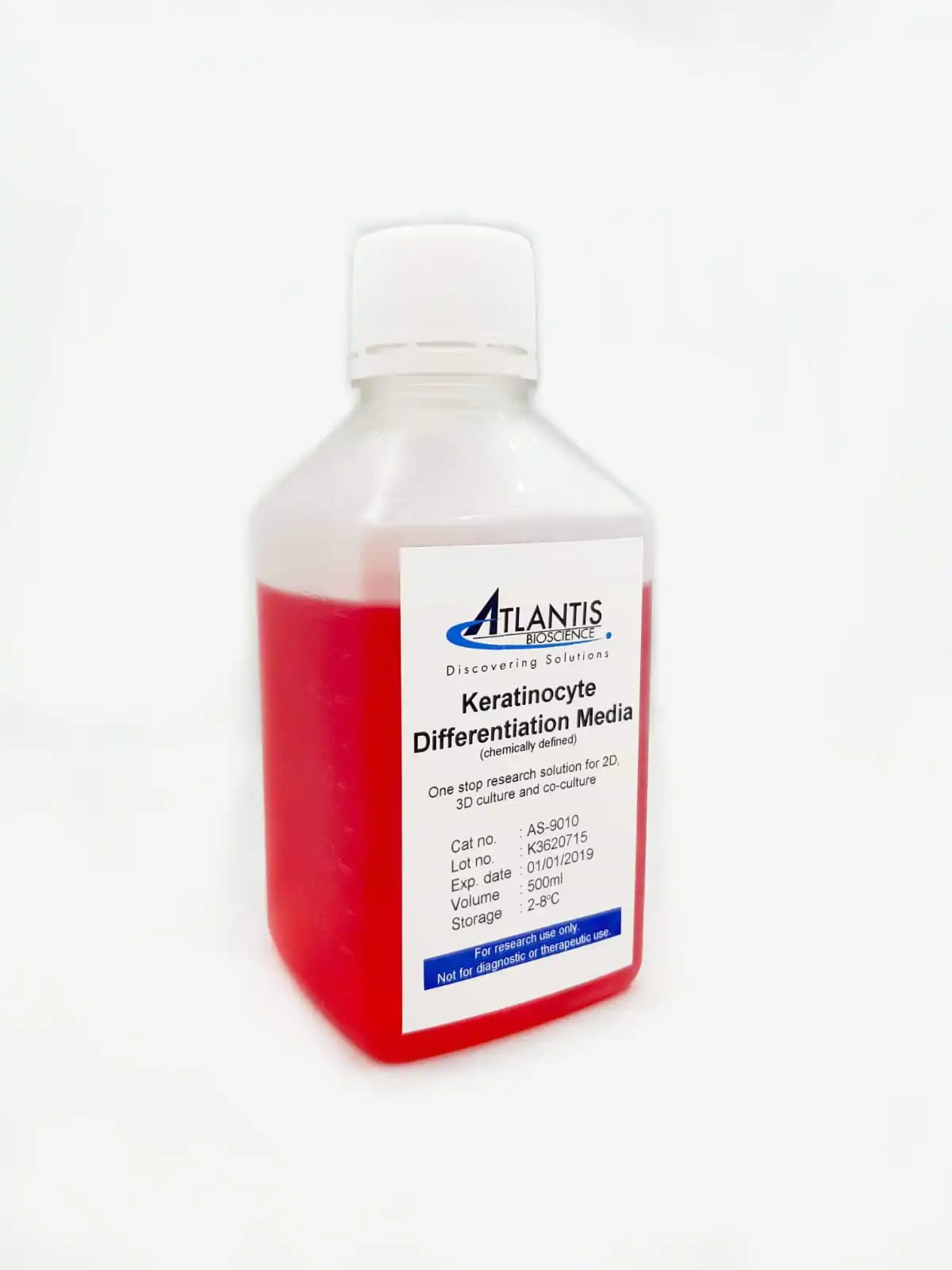

A chemically-defined serum-free media suitable for 3D organotypic Skin culture, 3D Keratinocyte collagen/airlift culture to allow for proper stratification of primary and immortalised keratinocyte.
It supports both the growth and differentiation of keratinocyte from the start of the culturing procedure to the end of the experimentation. This medium is also suitable to support the growth of skin cells embedded in Extracellular matrix such as fibroblast in collagen matrix.
Keratinocyte Differentiation Media (AS-9010) Used For 3D In-vitro Skin
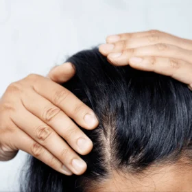
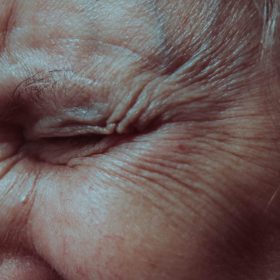
The EU has banned animal testing for cosmetics and their ingredients since 2013. A growing demand for in vitro skin models has been rising steadily to cater for both the safety testing (Skin irritation) and efficacy testing (functional test).
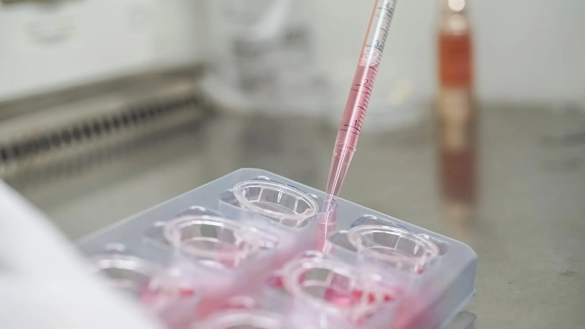
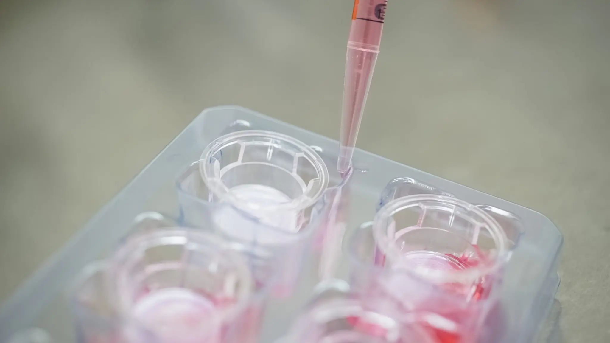
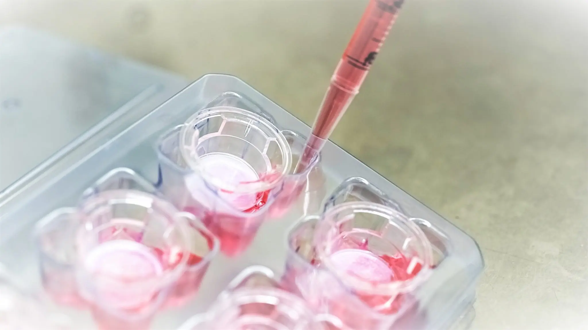
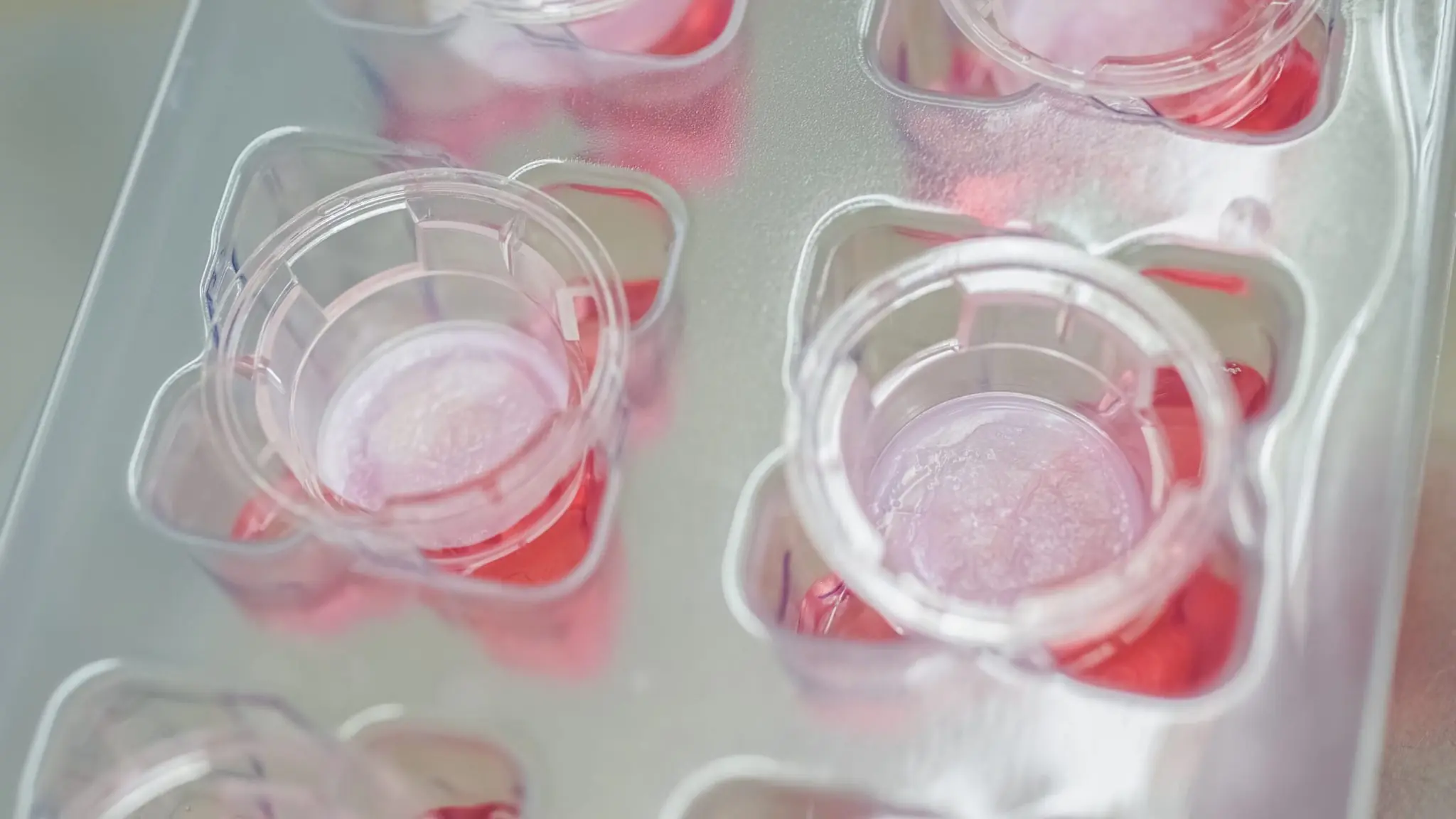
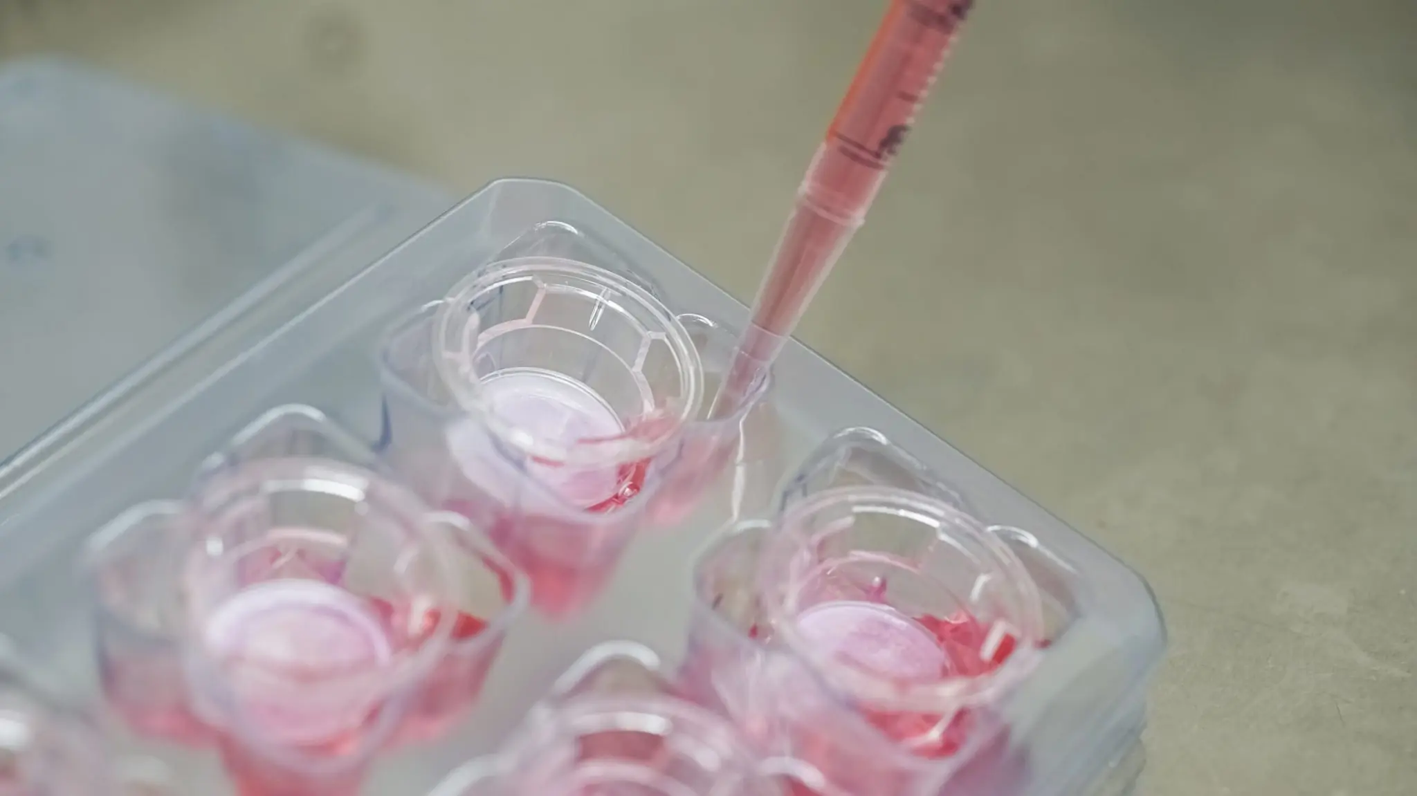
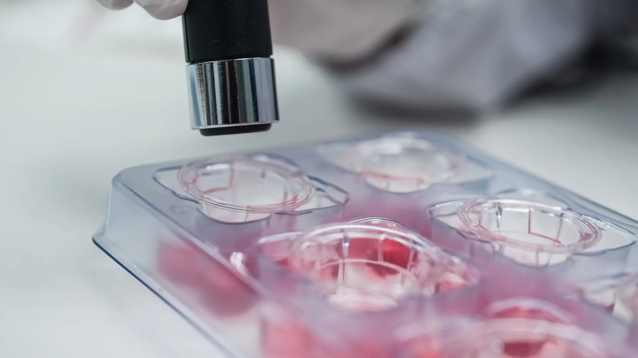
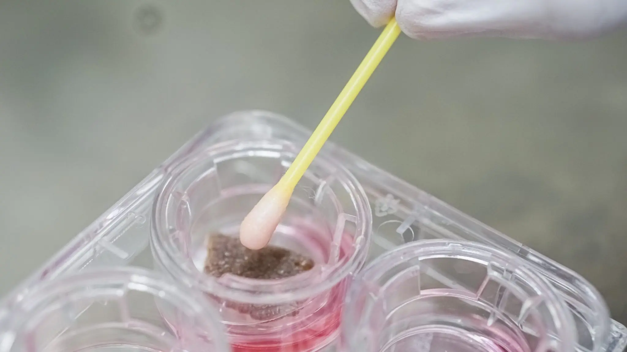
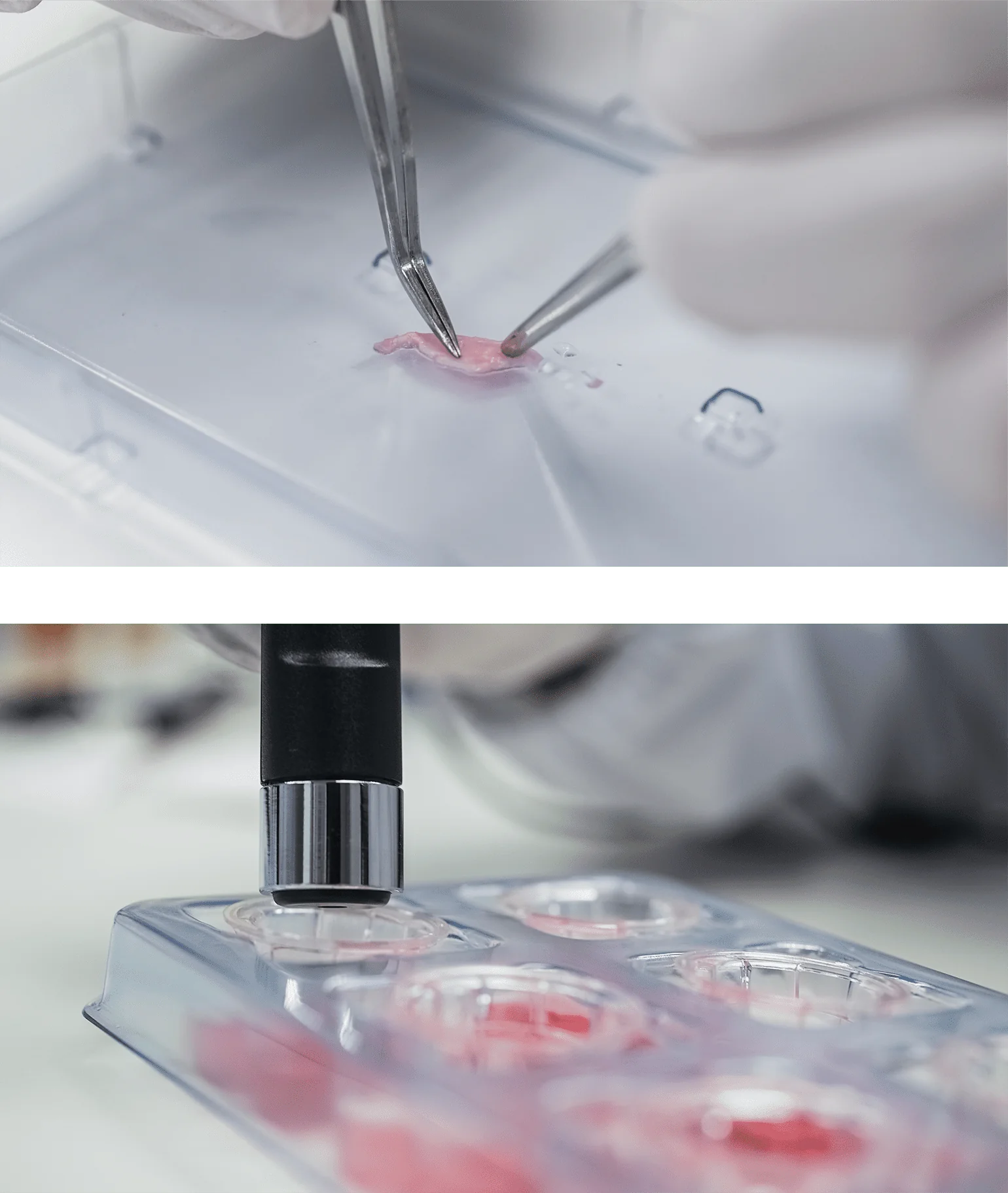
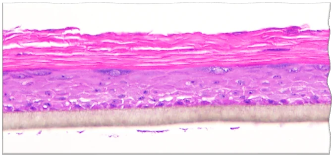
In the experiment, primary keratinocytes were seeded on a cultured insert to form a confluent layer. The confluent layer of cells is lifted to an air-liquid interface for 14 days to stimulates the keratinocytes to differentiate (stratification) and form a multi-layered epidermis that contains stratum corneum layers. The specialized medium primarily used consists of DMEM and Ham’s F12 in a specific ratio. It is chemically defined and serum-free to ensure a high reproducibility in result. Treatments can be topically applied to the skin model for a period of times. Histology such as H&E staining and other skin markers are commonly carried out. Gene expression and protein study are also alternative analysis method.
Materials:

Contact our Customer Care, Sales & Scientific Assistance
Consult and asked questions about our products & services
Documentation of Technical & Safety Data Sheet, Guides and more...
We gladly support you by keeping you updated on our latest products and the developments around our services.
362 Upper Paya Lebar Rd, #07-15,
Singapore 534963
© 2015-2023 Atlantis Bioscience Pte Ltd. All rights reserved. Co Reg No: 201539516N