Extracellular vesicles (EVs) are minuscule, membrane-bound structures that cells release into their environment. These vesicles act as cellular couriers, transporting proteins, DNA, and RNA molecules to other cells. Over the last decade, EVs have garnered significant attention due to their potential as natural carriers for complex cellular components and their involvement in various physiological and pathological processes.
Why EV Labeling Matters?
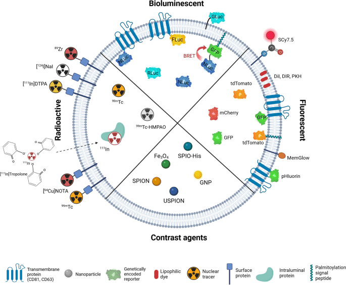
A multitude of in vitro and in vivo techniques are employed to delve into the biological functions of EVs. These include flow cytometry, confocal microscopy, and in vivo fluorescence detection methods. These techniques often rely on labelling EVs to facilitate their detection and analysis. Labelling strategies encompass optical (fluorescence, bioluminescence), nuclear, and magnetic resonance imaging (MRI) tracers, as illustrated in Figure 1. These advanced methods enable comprehensive insights into EV functions, interactions, and roles in physiological and pathological processes.
Figure 1: Strategies for labelling of exogenous extracellular vesicles.
Fluorescent Dyes for EV Staining: Current Approaches and Limitations
Detecting and characterising small EVs poses a significant challenge in the field. Flow cytometry, however, is frequently utilised for its ability to perform rapid, high-throughput, multiparameter analysis. Various fluorescent probes are commonly employed to detect small EVs using flow cytometry, including:
1. Antibody-Based EV Stains
These dyes exploit specific interactions with surface markers on small EVs, allowing for precise and targeted detection. These dyes are conjugated to antibodies that recognise and bind to distinct proteins on the EV membrane, facilitating their identification and characterisation. Notable surface markers include the tetraspanins CD9, CD63, and CD81. These proteins are highly enriched on the surface of EVs and are commonly used as markers for their detection. Antibody-based dyes targeting these tetraspanins provide specificity and sensitivity enabling the detailed analysis of EV populations, including their origin, composition, and potential functions. This targeted approach enhances the accuracy of flow cytometry in EV research, providing valuable insights into their roles in various physiological and pathological processes.
2. Membrane-Permeable EV Stains
Membrane-permeable dyes, such as carboxyfluorescein diacetate succinimidyl ester (CFSE), play a critical role in labelling and tracking EVs. CFSE is a lipophilic fluorescent dye that passively diffuses into cells and becomes highly fluorescent when intracellular esterases cleave its acetate groups. This reaction results in the dye covalently binding to amine groups in intracellular proteins, producing a stable and intense fluorescence signal. However, antigen expression and intracellular protein content vary depending on the cellular origin, leading to heterogeneity within a small EV population. Consequently, CFSE labelling may not be optimal for comparison across different samples. Additionally, the dye can leak from labelled cells, staining the surrounding environment and creating background noise that can obscure EV signals.
3. Lipophilic EV Stains
A variety of commercial lipid dyes are available, including PKH dyes and carbocyanine dyes (such as DiD, DiR, DiI). PKH dyes are highly fluorescent cell linkers that integrate into the cell membrane’s lipid bilayer. Common examples include PKH26 (red) and PKH67 (green). Although PKH dyes are widely used for labeling small EVs, there is growing consensus that they are unsuitable because they cause significant aggregation and alter vesicle size, and their uptake by cells produces false positives that compromise the reliability of experimental results. Studies have shown that PKH dyes can bind non-specifically to other cellular components or form free dye aggregates, creating background noise and complicating the identification of labelled EVs. PKH dyes might also affect membrane–membrane fusion, fluidity of membrane proteins, membrane stiffness and EV size.
Carbocyanine dyes, such as DiO and DiI, are another class of lipophilic dyes used for EV labelling. These dyes are known for their bright fluorescence and compatibility with various imaging techniques. However, these dyes suffer from poor solubility, leading to the formation of aggregates and fluorescent micelles that can easily be misinterpreted as EVs. This not only reduces labeling accuracy but also interferes with flow cytometry and fluorescence nanoparticle tracking analysis (fNTA), resulting in misleading data.
While lipophilic dyes like PKH and carbocyanine dyes are valuable tools for EV labelling, their limitations necessitate careful consideration and experimental design. Researchers must account for potential non-specific binding, aggregate formation, and impacts on membrane properties to ensure accurate and reliable EV detection. Exploring alternative dyes and labelling techniques may help mitigate these issues and enhance the precision of EV studies.
ExoBrite™: Next-Generation Dyes Optimized for EVs
Maintaining the integrity of EVs while ensuring accurate fluorescent labelling is paramount for reliable analysis. For extracellular vesicle membrane staining, modern purpose-built dyes such as Biotium’s ExoBrite™ seriesovercome these limitations by offering optimized chemistry specifically designed for EV research. Biotium has revolutionised EV labelling with their groundbreaking membrane dyes, offering a remarkable improvement over conventional methods.
ExoBrite™ True EV Membrane Stains – Best Pan-EV Coverage
ExoBrite™ True EV Membrane Stain represent a breakthrough in EV labeling technology, specifically optimized for extracellular vesicles. These stains provide higher, near-complete coverage of EVs within a sample, ensuring efficient labeling with minimal aggregation.
Unlike traditional dyes such as PKH, DiO, DiI, and DiD — which often form aggregates, distort vesicle size, and produce false positives — ExoBrite stains deliver bright, specific signals that can be clearly distinguished from nonspecific particles. Crucially, they are designed to address aggregation issues, allowing for clear and reproducible differentiation of EVs from background during flow cytometry detection and other analytical workflows. Analysis by fNTA showed ExoBrite™ True EV Membrane Stains detected ~96% of all EVs in a sample, compared to ~32% with PKH.
They are fully compatible with antibody co-staining, enabling researchers to combine membrane staining with surface marker phenotyping for deeper biological insights. For rigorous workflows, ExoBrite also aligns with best practices in EV methodology. When used with controls such as dye-only samples and detergent lysis assays, researchers can confirm that fluorescence originates from lipid-bound EVs, not free dye micelles or contaminants.
Customer Testimonial – NTU Researcher “ExoBrite stains EV membranes very effectively and provides clear visualization. It works especially well after SEC column purification, giving confidence that isolated EVs are being detected. Even with just 1 µL of stain, the signal is sharp and reliable.”
This makes ExoBrite staining highly reliable for flow cytometry, fNTA, and advanced imaging, providing reproducible, publication-quality EV analysis and advancing the precision of EV research.
ExoBrite™ EV Surface Stains
Biotium’s ExoBrite™ EV Surface Stains provide a targeted approach to labeling extracellular vesicles by binding to specific EV membrane surface. Each stain offers unique advantages depending on the biological question:
N-acetylglucosamine and sialic acid residues on EV surfaces
Bright signals; broad compatibility across diverse EV sources; suitable for purified or bead-bound EVs
General labeling of glycosylated EV subpopulations
Together, these surface-target specific EV stains expand the analytical toolkit for researchers by enabling selective labeling of biologically relevant EV membrane biomarkers. By minimizing aggregation compared to conventional dyes, ExoBrite™ EV Surface Stains provide clear and distinct signals for both purified and bead-bound EVs. This advanced technology supports accurate identification and characterization of EV subpopulations, offering researchers reproducible, high-resolution data and unlocking new opportunities in EV biology and biomedical research.
ExoBrite™ STORM CTB EV Stains for Super-Resolution Imaging
For researchers seeking advanced imaging capabilities, ExoBrite™ STORM CTB EV Stains offer unparalleled performance. These specialised fluorescent conjugates of CTB bind specifically to GM1 gangliosides on the surface of lipid rafts and EVs, enabling high-resolution STORM imaging. Incorporating STORM-validated CF® Dyes, these stains deliver exceptional clarity and sensitivity, even in the most demanding imaging conditions. Unlike traditional lipophilic dyes, ExoBrite™ STORM CTB EV Stains exhibit minimal background aggregation, ensuring precise identification and analysis of EVs with unprecedented accuracy.
The introduction of innovative dyes like ExoBrite™ by Biotium represents a significant leap forward in EV labelling and imaging technology. By overcoming the limitations of traditional dyes such as PKH and carbocyanine, these advancements provide researchers with reliable and precise tools for studying EV biology. These groundbreaking technologies not only enhance our understanding of EVs but also hold immense promise for their applications in medical research.
Antibody Conjugates for CD9, CD63, CD81
Biotium’s ExoBrite™ Antibody Conjugates are designed to provide researchers with highly specific tools for detecting canonical EV membrane biomarkers. These reagents target the tetraspanins CD9, CD63, and CD81, which are the most widely recognized markers for EV identification and characterization. By using optimized formulations, ExoBrite antibodies deliver brighter signals, reduced aggregation, and superior signal-to-noise, making them reliable across multiple EV analysis platforms.
Validated for robust detection; formulated in proprietary buffer to reduce antibody aggregation; brighter EV staining with optimal signal-to-noise; available as single-color cocktails (broad coverage) and 3-color cocktails (multiplexed phenotyping)
Conjugated to STORM-validated CF® Dyes for high-resolution EV visualization
These antibody-based tools ensure high specificity and minimal background, enabling accurate characterization of EV populations across flow cytometry, Western blot, and super-resolution microscopy workflows.
Best Practices for Reliable EV Labeling
To ensure reliable and publication-quality EV data, researchers should adopt rigorous controls and purification methods alongside their staining workflow:
Use dye-only controls: Detect and account for background fluorescence from free dye aggregates.
Apply detergent lysis controls: Confirm that fluorescence signals originate from lipid-bound EVs rather than contaminants.
Purify EVs before staining: Biofluids such as plasma contain lipoproteins that vastly outnumber EVs and can also be stained by lipophilic dyes. Proper purification (e.g., size exclusion chromatography) before staining is essential to remove these contaminants and improve labeling specificity.
Perform post-staining cleanup: After labeling, additional cleanup steps such as SEC columns or spin filters can be used to remove unbound dye and reduce background noise.
Balance sensitivity with specificity: Pan-EV membrane stains offer broad coverage, while surface-targeted stains or antibody conjugates allow precise analysis of EV subpopulations.
By integrating these best practices with optimized reagents such as ExoBrite™, researchers can achieve both technical rigor and biological accuracy, ensuring confidence in downstream EV analysis.
Reliable EV labeling is critical for accurate detection and characterization. Conventional dyes such as PKH and carbocyanine are increasingly discouraged due to artifacts and misleading signals, while purpose-built chemistries now offer reproducible, high-resolution results across flow cytometry, fNTA, Western blotting, and super-resolution imaging. Among these innovations, Biotium’s ExoBrite™ series provides optimized stains and antibody conjugates that align with best practices and enable researchers to generate data they can trust.
To explore these innovations firsthand, reach out to Atlantis Biosciencefor the latest promotions and product samples from Biotium.
References:
Bao, C., Xiang, H., Chen, Q., Zhao, Y., Gao, Q., Huang, F., & Mao, L. (2023). A Review of Labeling Approaches Used in Small Extracellular Vesicles Tracing and Imaging. International journal of nanomedicine, 18, 4567–4588. https://doi.org/10.2147/IJN.S416131
Boudna, M., Campos, A.D., Vychytilova-Faltejskova, P. et al. Strategies for labelling of exogenous and endogenous extracellular vesicles and their application for in vitro and in vivo functional studies. Cell Commun Signal 22, 171 (2024). https://doi.org/10.1186/s12964-024-01548-3
Dehghani, M., Gulvin, S.M., Flax, J. et al. Systematic Evaluation of PKH Labelling on Extracellular Vesicle Size by Nanoparticle Tracking Analysis. Sci Rep10, 9533 (2020). https://doi.org/10.1038/s41598-020-66434-7
Morales-Kastresana, A., Telford, B., Musich, T.A. et al. Labeling Extracellular Vesicles for Nanoscale Flow Cytometry. Sci Rep 7, 1878 (2017). https://doi.org/10.1038/s41598-017-01731-2
Nolan JP. Flow Cytometry of Extracellular Vesicles: Potential, Pitfalls, and Prospects. Curr Protoc Cytom. 2015;73:13.14.1-13.14.16. Published 2015 Jul 1. https://doi.org/10.1002/0471142956.cy1314s73
Pužar Dominkuš P, Stenovec M, Sitar S, et al. PKH26 labeling of extracellular vesicles: Characterization and cellular internalization of contaminating PKH26 nanoparticles. Biochim Biophys Acta Biomembr. 2018;1860(6):1350-1361. https://doi.org/10.1016/j.bbamem.2018.03.013
Simonsen JB. Pitfalls associated with lipophilic fluorophore staining of extracellular vesicles for uptake studies. J Extracell Vesicles. 2019;8(1):1582237. Published 2019 Feb 20. https://doi.org/10.1080/20013078.2019.1582237
Zhou C, Cox-Vázquez SJ, Chia GWN, et al. Water-soluble extracellular vesicle probes based on conjugated oligoelectrolytes. Sci Adv. 2023;9(2):eade2996. https://doi.org/10.1126/sciadv.ade2996
Liquid biopsy analysis has emerged as a groundbreaking tool in precision medicine, offering a minimally invasive way to detect and monitor diseases through biomarkers found
HOW CAN WE HELP YOU?Our specialists are to help you find the best product for your application. We will be happy to help you find the right product for the job.