🔥 Don’t Miss Out on our Blind Box Giveaway with a minimum spend of $280!🔥 Don’t Miss Out on our Blind Box Giveaway with a minimum spend of $280!🔥 Don’t Miss Out on our Blind Box Giveaway with a minimum spend of $280!
🔥 Don’t Miss Out on our Blind Box Giveaway with a minimum spend of $280!🔥 Don’t Miss Out on our Blind Box Giveaway with a minimum spend of $280!🔥 Don’t Miss Out on our Blind Box Giveaway with a minimum spend of $280!
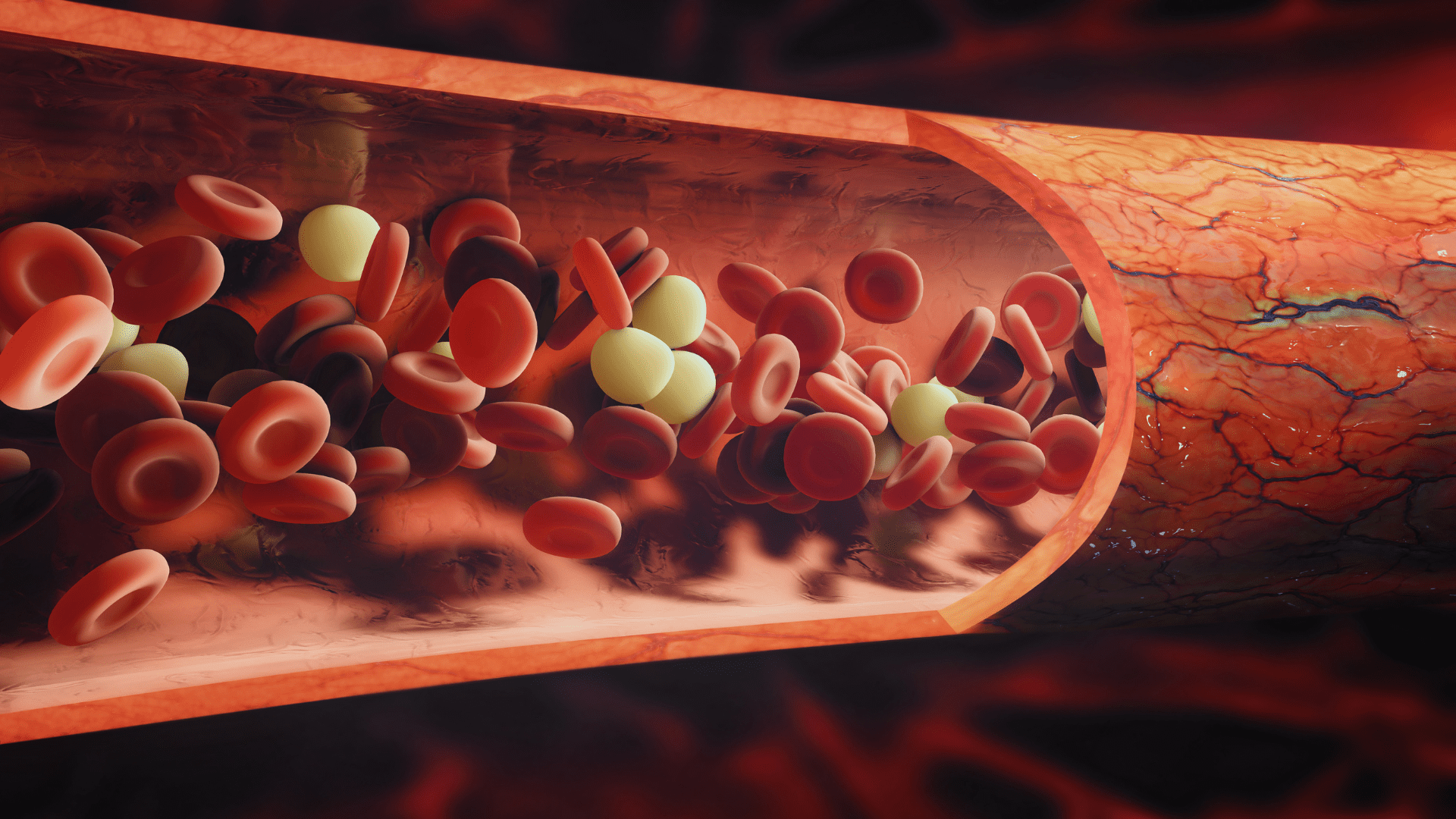
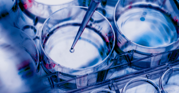
Shear stress is a mechanical force that acts parallel to a surface, exerted by a fluid as it flows over or through that surface. Various cell types experience shear stress in vivo, including kidney and lung epithelial cells, as well as endothelial cells that line blood vessels and lymphatic vessels.
How is shear stress measured?
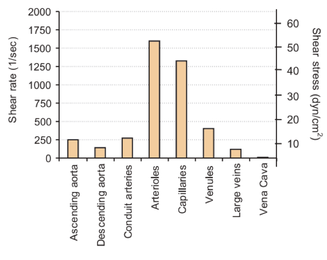
The magnitude of shear stress is measured in dyne/cm² and varies depending on the type of vessel and tissue involved (refer to Figure 1).
Figure 1: Shear stress values in various vessels. (Lipowsky et al., 1968)
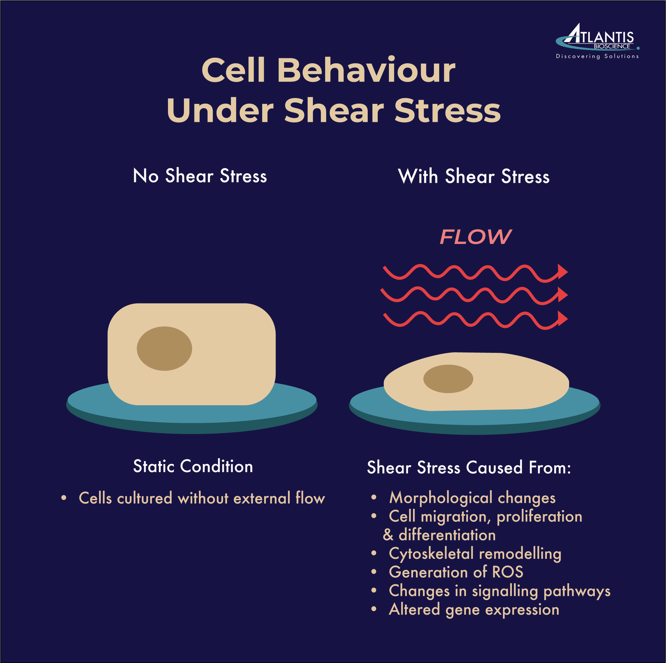
Shear stress plays a crucial role in numerous biological processes. It significantly influences cell behaviour, encompassing aspects such as cell proliferation, migration, and differentiation. Additionally, shear stress triggers intracellular signalling pathways that regulate gene expression and cellular responses (see Figure 2). For example, in the endothelial cells, shear stress stimulated the production of nitric oxide by the endothelial cells to help relax and widen blood vessels, promoting better blood flow and reducing vascular resistance. Moreover, shear stress activates mechanosensitive signalling pathways, such as the activation of transcription factors like AP-1 and NF-κB, which play a role in regulating the expression of genes involved in inflammation, vascular remodelling, and thrombosis.
Figure 2: Cell behaviour under shear stress
In conventional in vitro experiments, cells are often cultured without external flow, resulting in static conditions that do not replicate the dynamic and physiological environment found in vivo. In contrast, culturing cells under flow conditions in vitro can simulate this mechanical stimulus and induce a more physiologically relevant behaviour.
Microfluidic devices and Organ-on-chip are powerful tools that enable cell culture under perfusion with liquid culture media. By incorporating fluid flow and controlled shear stress levels, these systems can better mimic physiological conditions, and provide more accurate models for studying organ function, disease mechanisms, and drug responses.
6 objectives of scientists studying shear stress on cell culture
Here are some specific applications for investigating the effect of shear stress on cell culture:
Vascular research: Long-term culture of endothelial cells under mechanical stress to study the physiology and responses of these cells.
Air-liquid interface (ALI) research: Creating a model that mimics the endothelium-epithelium barrier by culturing 2D/3D epithelial cells in the upper well and seeding endothelial cells in the perfusable channel underneath. This setup is commonly used for studying barriers like the Blood-Brain Barrier (BBB) and those found in the gut, skin, lung, and other organs.
Coculture research: Investigating the crosstalk between endothelium and epithelium under flow conditions to study the interactions and responses of different organs such as kidney, liver, gut, or heart.
Hypoxia studies: Combining 3D and 2D cell cultures in a hypoxic environment where oxygen is supplied to the cells via the medium flowing in one of the channels. This allows the study of cellular responses to low oxygen levels.
Chemotactic migration studies: Applying nutrient, oxygen, or drug gradients to study cell migration under these conditions, providing insights into cell behaviour in chemotactic environments.
Rolling and adhesion assays: Investigating the effect of circulating particles, such as bacteria, immune responses, or circulating tumour cells, on cell rolling and adhesion behaviours under flow conditions.
By utilising microfluidic devices and Organ-on-chip systems, researchers can create more physiologically relevant cell culture models and gain deeper insights into cellular responses under the influence of shear stress and fluid flow. These approaches provide valuable tools for studying various biological processes and disease mechanisms.
References:
Nakajima H, Mochizuki N. Flow pattern-dependent endothelial cell responses through transcriptional regulation. Cell Cycle. 2017;16(20):1893-1901. doi:10.1080/15384101.2017.1364324
Papaioannou TG, Stefanadis C. Vascular wall shear stress: basic principles and methods. Hellenic J Cardiol. 2005;46(1):9-15.
Whitmore RL: Rheology of the circulation. New York: Pergamon Press, 1968.
Singapore has emerged as a global hub for innovation and entrepreneurship, particularly in the life sciences sector. The city-state’s strategic position, robust infrastructure, and supportive
The Future is Flexible: Summary Substrate stiffness serves as a critical determinant of MSC behaviour, exerting profound effects on adhesion, proliferation, migration, and differentiation. Understanding
HOW CAN WE HELP YOU?Our specialists are to help you find the best product for your application. We will be happy to help you find the right product for the job.