- Your cart is empty
- Continue Shopping


Modern biology has long relied on techniques that analyse cells in isolation. While bulk and single cell sequencing have provided deep molecular insights, they often miss a crucial dimension: spatial context. In spatial biology, preserving tissue architecture is essential for understanding how cells interact within their native microenvironment.
TSA-based multiplex immunofluorescence (mIF) addresses this limitation by enabling high sensitivity detection of multiple protein targets within intact tissue sections. By combining TSA with sequential staining strategies, researchers can map functional protein expression while maintaining spatial relationships between cells.
Traditional techniques such as bulk sequencing and even single-cell sequencing typically require tissue dissociation. While these methods reveal valuable information about gene or protein expression, they disrupt the spatial relationships between cells.
However, in many biological processes, location is as important as molecular identity. For example:
Without spatial information, we lose sight of cell–cell interactions, functional niches, and the microenvironmental influences that drive disease progression. TSA-based mIF within a spatial biology framework enables high sensitivity protein mapping while preserving these critical spatial relationships.

Multiplex immunofluorescence (mIF) is a cornerstone technique in spatial biology, enabling detection of multiple protein markers on a single tissue section while preserving tissue architecture. In many high-sensitivity workflows, mIF incorporates Tyramide Signal Amplification, often referred to as TSA-based mIF, to enhance signal intensity and enable reliable detection of low-abundance protein targets.
While mIF provides high-resolution protein-level insight, spatial biology encompasses a broader toolkit often referred to as spatial omics. These technologies enable molecular mapping at scale within intact tissues, including:
Together, these approaches provide complementary layers of information. Spatial transcriptomics supports unbiased discovery, whereas TSA-based mIF delivers high-sensitivity protein validation within preserved tissue architecture. This multi-layered framework allows researchers to interpret molecular programmes in the context of cellular organisation and microenvironmental structure.
Careful experimental design is critical for generating reliable spatial biology data, particularly when implementing TSA-based mIF workflows.

Selecting the right spatial biology platform is often the most challenging step in experimental design. It is tempting to pursue the highest plex method available, but the most appropriate technology is the one that answers your specific biological question with sufficient resolution, sensitivity, throughput, and reproducibility.
While this guide focuses on TSA-based multiplex immunofluorescence (mIF), it is essential to understand how it compares to other leading spatial technologies. The table below outlines common scenarios to help determine when TSA-mIF is the optimal choice—and when alternatives may be better suited.
| Technology | Best Used For | Plex Capacity | Sensitivity | Why Choose TSA-mIF Instead? |
| TSA-based mIF | Validating 4–10 markers; high-sensitivity detection of “dim” targets. | 4–10 | Highest (10-100x boost) | Cost-effective. Uses standard lab microscopes; covalent signal allows stripping without data loss. |
| PhenoCycler (CODEX) | High-plex discovery; mapping 40+ cell types in a single tissue. | 40–60+ | Moderate | Less Complexity. PhenoCycler requires specialised fluidics and expensive barcodes; TSA is better for low-abundance proteins. |
| Imaging Mass Cytometry (IMC) | Autofluorescence-heavy tissues (Lung/Liver); absolute quantification. | 40+ | Low to Moderate | Higher Resolution. IMC resolution is often limited to 1µm; TSA-mIF offers crisp subcellular detail and much higher signal. |
| Spatial | Whole-transcriptome discovery; RNA-focused questions. | 1,000–10,000+ | Variable | Protein is King. RNA levels don’t always equal protein levels. TSA validates the “functional” protein expression in situ. |
TSA-based mIF is the workhorse for hypothesis-driven protein imaging. It is the only method listed that combines (a) FFPE compatibility, (b) single-molecule sensitivity, (c) standard laboratory equipment, and (d) established utility in high-impact translational research.
If your experiment requires 40+ markers or unbiased discovery, consider CODEX or spatial transcriptomics. If you need to visualise 4–10 carefully selected proteins with high sensitivity and reproducibility, TSA-mIF is the superior choice.
As we move toward higher-plex imaging, traditional staining methods often struggle with weak signals. Tyramide Signal Amplification (TSA) has emerged as a key technology to bridge this gap.
Note on scope: Spatial omics encompasses RNA, protein, and metabolite mapping. This section focuses specifically on multiplexed protein imaging, where sensitivity is often the limiting factor. While spatial transcriptomics captures the blueprint of gene expression, spatial proteomics reveals the functional state of cells—post-translational modifications, activated signalling pathways, and therapeutic target engagement. TSA is the critical enabling chemistry for this domain.
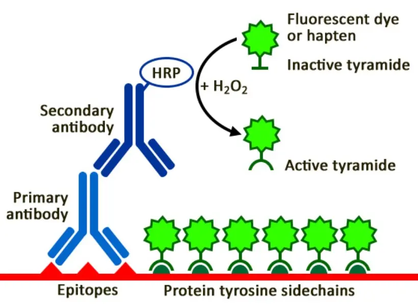
TSA is an enzyme-mediated detection method that uses Horseradish Peroxidase (HRP) to catalyse the deposition of labelled tyramide molecules. These reactive radicals bind covalently to nearby tyrosine residues on the tissue.
The result? A signal that is 10–100 times stronger than conventional IHC. Because the bond is covalent, the fluorescent signal stays “locked” in place even if the primary antibodies are stripped away to make room for the next round of markers.
Choosing the right amplification chemistry is the most critical decision in panel development. While several commercial options exist, researchers prioritise brightness, photostability, and ease of automation.
When evaluating the best TSA kits, Biotium offers standout tailored for specific research needs:

Compared with commonly used tyramide systems such as Opal and Aluora, TyraMax™ is engineered to deliver higher fluorescence intensity and improved resistance to photobleaching during repeated imaging and stripping cycles. This enhanced performance supports clearer detection of low-abundance protein targets and more stable signal retention in high-plex spatial imaging applications.
Importantly, TyraMax™ reagents remain stable in amplification buffer for up to 24 hours, making them well suited for automated staining platforms where reagent stability and consistency are essential for reproducible multiplex workflows.
By selecting kits that offer high signal-to-noise ratios and chemical stability, labs can significantly reduce the “trial and error” often associated with high-plex TSA workflows.
Despite its strengths, TSA-based multiplexing presents practical challenges:
Strategies to overcome these challenges include:
Well-optimised TSA workflows enable reliable, reproducible, and high-resolution spatial maps of protein expression within tissues.
The future of spatial biology lies in integration. Advances in AI-driven image analysis are enabling correlation of a cell’s genetic state with its physical location and neighbouring proteomic profile within intact tissue. As TSA-based multiplex immunofluorescence workflows become more automated and higher-throughput, high-sensitivity spatial protein mapping is increasingly positioned to support translational research and clinical diagnostics.

Spatial biology is transforming our understanding of life by preserving the “neighbourhood” in which cells live and work. By leveraging the sensitivity of TSA and the power of multiplex imaging, researchers are finally seeing the full picture—accelerating discoveries that will define the next generation of precision medicine.
Bressan D, Battistoni G, Hannon GJ. The dawn of spatial omics. Science. 2023 Aug 4;381(6657):eabq4964. doi: 10.1126/science.abq4964.
Liu Y, Dai Y, Wang L. Spatial omics at the forefront: emerging technologies, analytical innovations, and clinical applications. Cancer Cell. 2026 Jan 12;44(1):24-49. doi: 10.1016/j.ccell.2025.12.009.
Moffitt JR, Lundberg E, Heyn H. The emerging landscape of spatial profiling technologies. Nat Rev Genet. 2022 Dec;23(12):741-759. doi: 10.1038/s41576-022-00515-3.
Radosevic-Robin N, Kossai M, Penault-Llorca F. New-generation technologies for spatial tissue analysis, indispensable tools for deciphering intratumor heterogeneity in the development of antibody-drug conjugates and radio-immunoconjugates for cancer treatment. Transl Breast Cancer Res. 2023 Sep 20;4:28. doi: 10.21037/tbcr-23-38.
Sheng W, Zhang C, Mohiuddin TM, Al-Rawe M, Zeppernick F, Falcone FH, Meinhold-Heerlein I, Hussain AF. Multiplex Immunofluorescence: A Powerful Tool in Cancer Immunotherapy. Int J Mol Sci. 2023 Feb 4;24(4):3086. doi: 10.3390/ijms24043086.
Stack EC, Wang C, Roman KA, Hoyt CC. Multiplexed immunohistochemistry, imaging, and quantitation: a review, with an assessment of Tyramide signal amplification, multispectral imaging and multiplex analysis. Methods. 2014 Nov;70(1):46-58. doi: 10.1016/j.ymeth.2014.08.016.
Tan WCC, Nerurkar SN, Cai HY, Ng HHM, Wu D, Wee YTF, Lim JCT, Yeong J, Lim TKH. Overview of multiplex immunohistochemistry/immunofluorescence techniques in the era of cancer immunotherapy. Cancer Commun (Lond). 2020 Apr;40(4):135-153. doi: 10.1002/cac2.12023.

Connect With Our Technical Specialist.

Request For A Quotation.
DON’T MISS OUR UPDATES.
FOLLOW US ON SOCIAL MEDIA!
![]()
![]()
![]()
Contact our Customer Care, Sales & Scientific Assistance
Consult and asked questions about our products & services
Documentation of Technical & Safety Data Sheet, Guides and more...