Extracellular vesicles (EVs) and exosomes are gaining increasing attention for their roles in diagnostics, therapeutics, and biomarker discovery. These tiny vesicles carry proteins, RNA, and other cargo from their parent cells, acting as natural messengers with functional impacts on recipient cells and playing a central role in cell-to-cell communication.
Exosomes are nano-sized EVs, typically 30–150 nanometres in diameter, that are secreted by most cell types and found in nearly all biological fluids — including plasma, serum, saliva, cerebrospinal fluid, urine, and cell culture media. They encapsulate diverse biomolecules from their cell of origin, such as nucleic acids, proteins, lipids, and metabolites, serving as natural carriers for intercellular communication. While exosomes were first thought to function primarily in cellular waste disposal, later studies revealed their crucial role in regulating physiological and pathological processes.
For instance, mesenchymal stem cell–derived exosomes (MSC-Exosomes) have been widely studied for their regenerative and therapeutic potential. Beyond regenerative medicine, exosomes are now explored as diagnostic and prognostic biomarkers for cancer, cardiovascular disorders, tuberculosis, and neurodegenerative diseases. Their intrinsic biocompatibility, low immunogenicity, and efficient cargo delivery also make them promising drug delivery systems, with ongoing studies investigating their applications in cancer and Alzheimer’s disease therapy.
Harnessing this potential, however, requires meticulous planning, reproducible workflows, and optimised analytical tools.
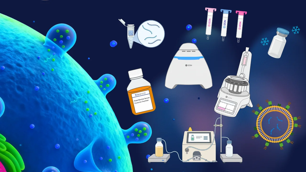
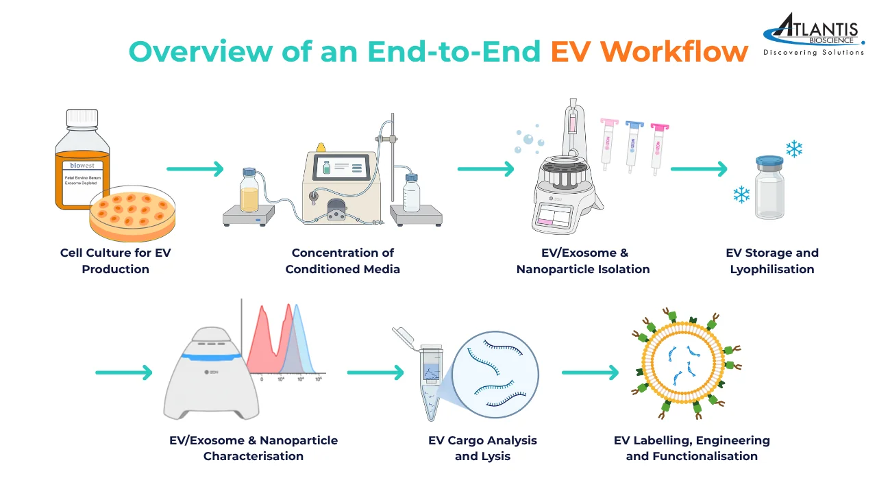
In this blog, we walk through a day in the EV lab, following a vesicle from culture through concentration, isolation, storage, characterisation, cargo analysis, and functionalisation—highlighting real lab experiences, best practices, and practical tips.
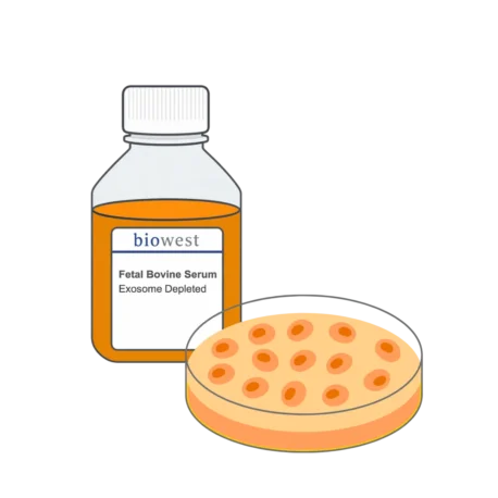
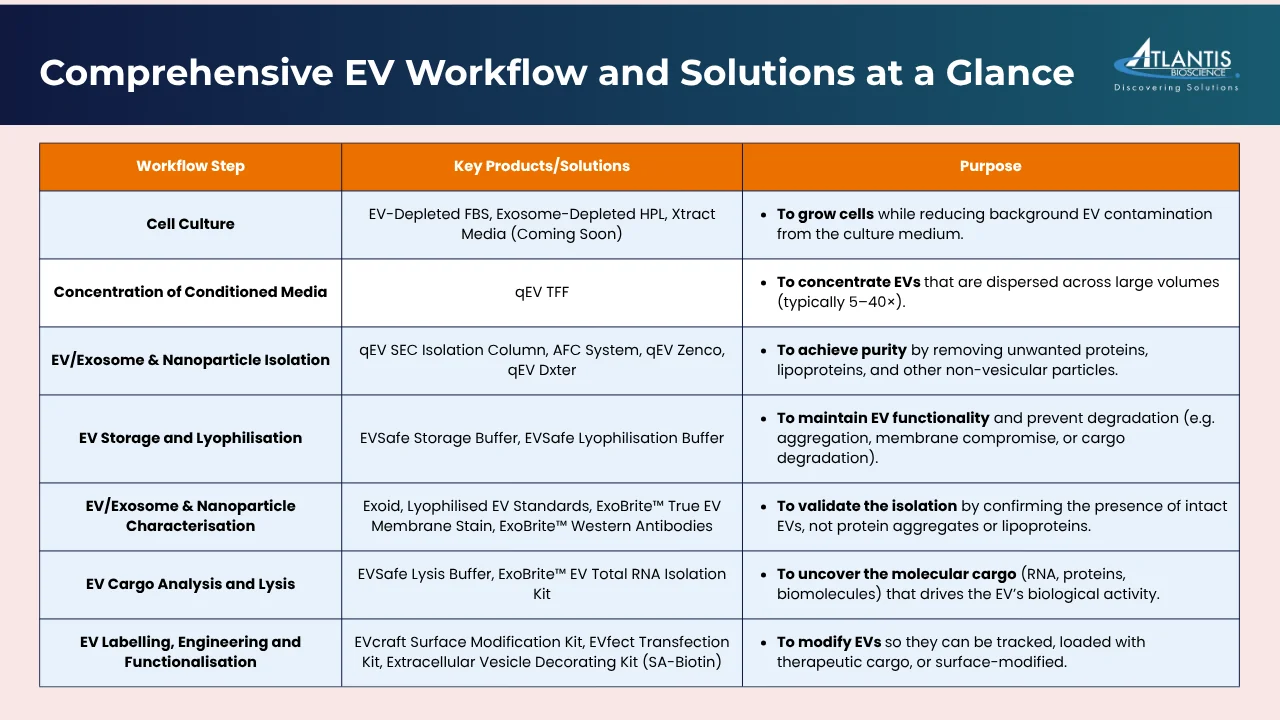
Step 1: Cell Culture for EV Production
The day begins at the incubator. Your cells are thriving, but there’s a hidden challenge: the culture medium itself can introduce EV contamination. Standard fetal bovine serum (FBS) contains bovine-derived EVs and non-EV particles like lipoproteins, which can obscure results or reduce apparent yield.
What happens in the lab:
You seed your mesenchymal stem cells (MSCs) or immune cells in EV-depleted media. The cells continue their normal secretory activity, releasing EVs into the conditioned media over several days.
During this period, it’s crucial to maintain optimal culture conditions—temperature, CO₂ levels, and cell density—as stressed cells can release apoptotic bodies, which are larger vesicles that may complicate downstream analyses.
Solutions:
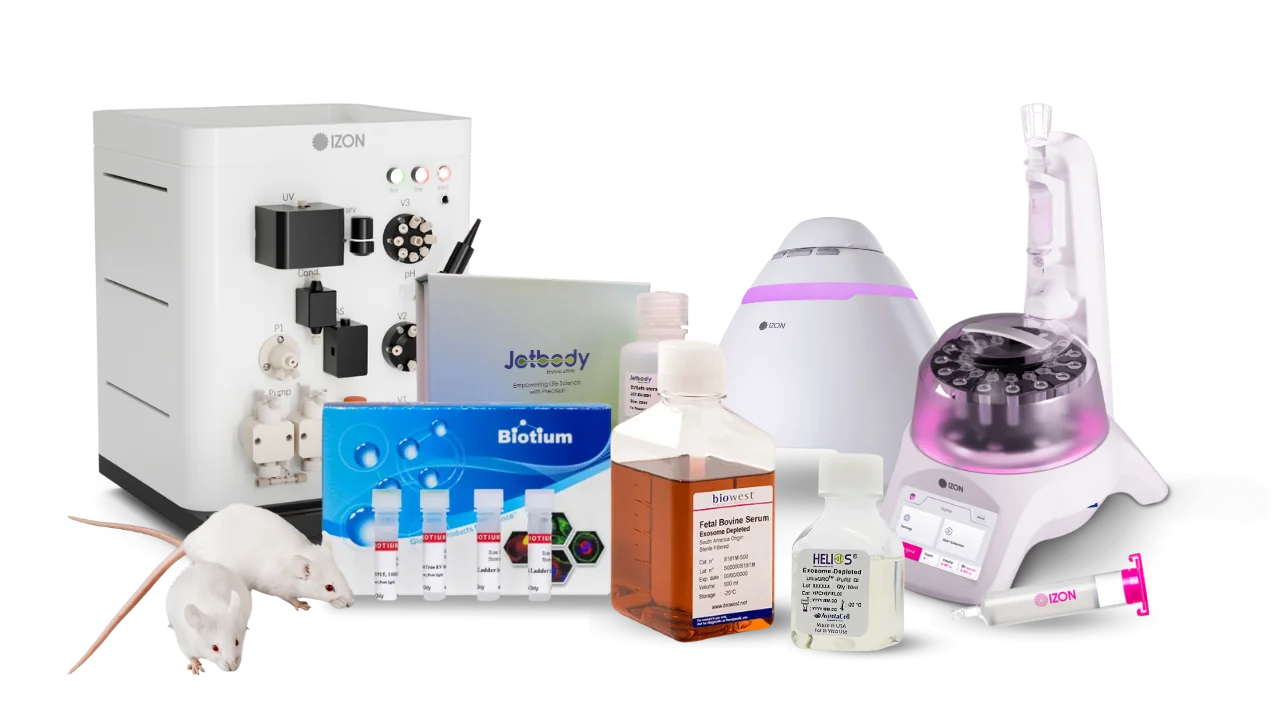
EV-Depleted FBS – reduces background EV contamination for serum-based cultures.
Exosome-Depleted HPL – human platelet lysate optimised to avoid exogenous vesicles.
Xtract Media (Coming Soon) – a chemically defined medium designed for high-yield EV production.
Tip
Pitfall
Pre-condition your media and confirm depletion of contaminating vesicles before starting experiments.
Over-confluent cell cultures can alter the therapeutic potential of MSCs due to contact-inhibited growth or contact-induced senescence.
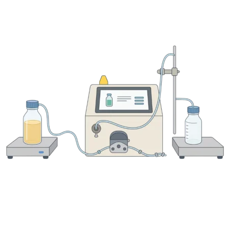
Step 2: Concentration of Conditioned Media for EV Isolation
After the culture period, your conditioned media is rich in EVs—but they are dispersed across large volumes, making isolation inefficient. In most EV workflows, samples are first concentrated (typically 5–40×) before purification because conditioned media are often too dilute to process directly. Concentration is therefore a crucial bridge between culture and isolation.
What happens in the lab:
You carefully transfer the conditioned media into the tangential flow filtration (TFF) system. The media circulates across semi-permeable membranes, allowing small molecules and water to pass through while retaining and concentrating the EVs.
Ideal for processing large volumes (e.g., >250 mL), TFF employs membranes with defined molecular weight cut-offs (MWCOs)—commonly 100 kDa, 300 kDa, or 750 kDa—depending on the desired purity and the characteristics of the vesicles.
Gentle handling throughout is essential; excessive pressure, high shear, or aggressive centrifugation can damage vesicle membranes or alter their surface properties.
Solution:
qEV TFF (Tangential Flow Filtration) – scalable and gentle concentration method that preserves EV integrity.
Tip
Pitfall
Monitor flow rate and pressure to avoid vesicle deformation.
Over-concentration can increase viscosity, leading to aggregation or losses during subsequent isolation.
Step 3: EV/Exosome & Nanoparticle Isolation
Purity is everything. After concentration with TFF, your sample is denser but still contains unwanted proteins, lipoproteins, and other non-vesicular particles. To obtain biologically relevant and reproducible data, these contaminants must be removed—this is where true EV isolation begins.
What happens in the lab:
Using size-exclusion chromatography (SEC), your concentrated sample is loaded onto a qEV column. As it flows through the packed resin under gentle gravity, larger EVs elute first while smaller proteins, lipoproteins, and other soluble contaminants are delayed.
Because qEV relies on gravity rather than pressure, the process is exceptionally gentle—preserving the vesicles’ morphology, membrane integrity, and biological function.
For researchers seeking higher consistency and reproducibility, the AFC system automates fraction collection from qEV columns. This ensures precise control over fraction timing and volume, minimizing user variability and simplifying the workflow.
For large-scale production, the qEV Zenco—an FPLC system integrated with qEV columns—enables automated, scalable EV isolation while maintaining the same gentle SEC principle.
Meanwhile, qEV Dxter offers high-throughput processing capabilities, allowing multiple samples to be isolated simultaneously in parallel instruments for core facilities or large research programs.
Solutions:
qEV SEC Isolation Column – manual or automated size-exclusion chromatography for high-purity EV isolation.
AFC System – automates fraction collection for consistent, reproducible EV purification and reduced manual handling.
qEV Zenco– FPLC-based automation for large-scale EV isolation.
qEV Dxter– high-throughput system for multi-sample processing across multiple columns.
Tip
Pitfall
Match your isolation method to your sample volume and throughput needs.
Skipping column equilibration or insufficient washing can introduce contaminants that compromise downstream characterisation.
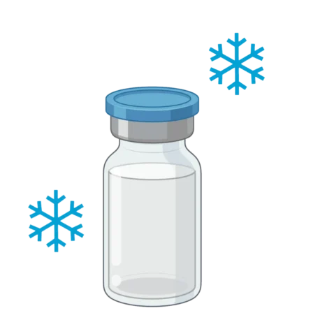
Step 4: EV Storage and Lyophilisation for Long-term Stability
After isolation, proper storage is essential to maintain EV functionality. Vesicles are delicate—improper conditions can compromise membrane integrity, cause aggregation, or degrade their RNA and protein cargo. Yet, storage remains one of the least standardised steps in EV workflows.
Typically, isolated EVs are resuspended in phosphate-buffered saline (PBS) and stored at −80 °C or lower, as refrigeration at 4 °C leads to loss of particle number, altered size distribution, and decreased expression of surface markers. Even at −80 °C, repeated freeze–thaw cycles can rupture vesicles and cause aggregation.
To address these challenges, researchers have turned to lyophilisation (freeze-drying). As highlighted in recent studies, lyophilised EVs—especially those stabilised in protective buffers—can retain their size, morphology, and biological function for extended periods, even at room temperature. This approach offers a more practical and cost-effective alternative for long-term EV preservation and transport without constant cold-chain dependency.
What happens in the lab:
You aliquot freshly isolated EVs into cryovials to minimise freeze–thaw cycles.
Some batches are lyophilised using EVSafe Lyophilisation Buffer, allowing long-term, ambient-temperature storage. When reconstituted, the EVs show minimal change in particle size or zeta potential—ready for downstream analyses or functional assays.
Solutions:
EVSafe Storage Buffer – maintains EV stability during freezing with >95% recovery. Prevents aggregation and preserves particle size distribution during storage at −80 °C.
EVSafe Lyophilisation Buffer– formulated for freeze-drying EVs while maintaining vesicle structure and function (~85% recovery). Enables easy reconstitution for downstream applications and stable storage at room temperature.
Tip
Pitfall
Always aliquot EVs into single-use volumes to prevent degradation from repeated thawing.
Freezing or thawing EVs without cryoprotectants can rupture vesicles and degrade RNA/protein cargo, significantly affecting assay reproducibility.
Isolation alone doesn’t confirm success. Characterisation validates what you’ve truly isolated—ensuring that your sample contains intact EVs rather than protein aggregates or lipoproteins. Proper characterisation is critical for reproducibility, comparability between studies, and ensuring that observed biological effects are indeed mediated by EVs.
Why it matters:
Physical and surface characterisation provides insight into size, concentration, zeta potential, and membrane composition—key parameters that influence EV stability, cellular uptake, and functional behaviour. Knowing the size distribution helps distinguish small EVs (exosomes, ~30–150 nm) from microvesicles or non-vesicular particles. Zeta potential reflects surface charge and colloidal stability, while particle concentration informs accurate dosing in functional assays.
What happens in the lab:
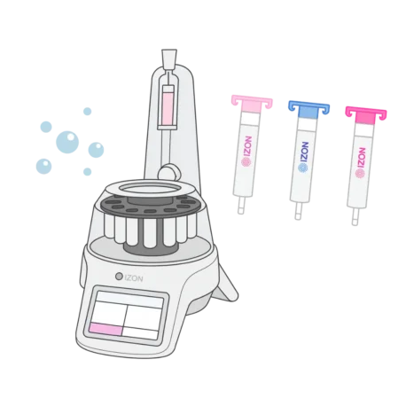
A fraction of isolated EVs is loaded onto the Exoid, which uses Tunable Resistive Pulse Sensing (TRPS) to measure each particle individually. This provides true particle-by-particle size, concentration, and zeta potential data, avoiding biases from aggregates or mixed populations common in Dynamic Light Scattering (DLS) or Nanoparticle Tracking Analysis (NTA).
Transmission Electron Microscopy (TEM) is used to visualise EV morphology, confirming the classic cup-shaped structure and verifying the absence of debris or protein aggregates.
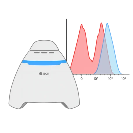
For surface profiling, flow cytometry is performed using ExoBrite™ True EV Membrane Stain. Unlike traditional lipophilic dyes such as Dil or PKH—which can induce EV aggregation —ExoBrite™ selectively stains intact EV membranes with minimal aggregation. When combined with ExoBrite™ CD marker–specific antibodies (e.g., CD63, CD81, CD9), it allows accurate detection of canonical EV markers and identification of subpopulations.
ExoBrite™ Western Antibodies provide optimised antibodies for EV protein marker detection, including CD9, CD63, and CD81, and Calnexin (negative control), producing clean, reproducible signals. This complements physical characterisation, offering molecular confirmation of EV identity.
Solutions:
Lyophilised EV Standards – reference materials from different cell sources for calibration and comparison.
Exoid (TRPS Technology) – precise measurement of particle size, concentration, and zeta potential, with single-particle resolution.
ExoBrite™ Western Antibodies – validated antibody conjugates for protein marker profiling with minimal background.
Tip
Pitfall
– Use orthogonal characterisation methods—TRPS, TEM, flow cytometry, and Western blot—to comprehensively understand your EV prep.
– ExoBrite™ reduces aggregation and background compared to traditional dyes, ensuring more accurate flow cytometry results.
– Over-reliance on a single technique, such as DLS or NTA, can misrepresent size or concentration, particularly in polydisperse samples.
– Skipping validation of surface markers may overlook heterogeneous subpopulations or contaminants, affecting downstream functional studies.
Step 6: EV Cargo Analysis and Lysis
The true story of EVs lies within. While isolation and physical characterisation confirm the presence of vesicles, it is their molecular cargo—RNA, proteins, and other biomolecules—that often drives their biological activity. Understanding what EVs carry is essential for uncovering their role in intercellular communication, identifying disease biomarkers, and developing EV-based therapeutics.
Why cargo analysis matters:
Biomarker discovery: EVs reflect the physiological state of their parent cells, making them valuable for liquid biopsies and disease monitoring. For example, tumour-derived EVs may carry oncogenic RNAs or proteins that serve as early indicators of cancer.
Therapeutic development: Many therapeutic strategies exploit EVs as natural delivery vehicles for drugs, RNA therapeutics, or signalling proteins. Characterising cargo ensures the vesicles contain the intended functional molecules.
Mechanistic studies: Profiling cargo helps understand EV-mediated cell signalling and intercellular communication in health and disease. Functional assays can reveal how EVs transfer RNA or proteins to recipient cells to induce biological effects.
What happens in the lab:
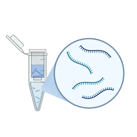
You gently lyse the EVs using a dedicated EVSafe Lysis Buffer, which disrupts the vesicle membrane while preserving the integrity of nucleic acids and proteins. Standard lysis reagents can be too harsh, leading to fragmentation or degradation of cargo. EV-specific buffers are formulated to maintain molecular stability while releasing internal content efficiently.
RNA analysis: Using the ExoBrite™ EV Total RNA Isolation Kit, RNA is extracted for next-generation sequencing (NGS), qPCR, or microarray studies, enabling detailed transcriptomic profiling of EV cargo.
Protein analysis: Proteins are quantified and characterised via Western blotting or mass spectrometry, allowing identification of signalling molecules, enzymes, or disease-associated proteins.
Functional assays: Reporter genes or cellular uptake assays can assess the delivery and activity of EV cargo in target cells, providing insight into biological effects and therapeutic potential.
EVSafe Lysis Buffer – gentle yet effective, formulated to lyse vesicles while maintaining cargo stability for downstream molecular and functional analyses.
Tip
Pitfall
– Always use EV-specific lysis buffers to preserve RNA and protein quality, especially for sensitive assays like NGS or mass spectrometry.
– Combine molecular profiling with functional assays to validate that the cargo is biologically active.
– Harsh lysis conditions can fragment RNA or denature proteins, compromising downstream analyses and obscuring functional insights.
– Skipping proper lysis or purification steps may leave membrane remnants that interfere with accurate cargo quantification.
Step 7: EV Labelling, Engineering and Functionalisation
Finally, your EVs are ready for advanced applications. Beyond their natural biological roles, EVs can be tracked, loaded with therapeutic cargo, or surface-modified to enhance targeting, delivery efficiency, and stability. Functionalisation enables EVs to become therapeutic and diagnostic tools, allowing researchers to direct them to specific tissues, protect cargo like RNA or drugs from degradation, and enable real-time tracking for diagnostics. This customisation unlocks capabilities far beyond what natural EVs can achieve alone.
What happens in the lab:
Using the EVfect Transfection Kit, you introduce therapeutic RNA—such as siRNA, miRNA, mRNA, or antisense oligonucleotides—into EVs. This allows targeted delivery of functional nucleic acids to recipient cells.
Decorate the EV surface using the Extracellular Vesicle Decorating Kit (SA-Biotin). This Sortase A–mediated enzymatic reaction enables precise and stable protein modification on the EV membrane, facilitating conjugation to targeting molecules.
With the EVcraft Surface Modification Kit, a wide range of surface molecules can be conjugated onto EVs:
Antibodies or nanobodies for antigen-specific cellular targeting.
Ligands such as agonists or cytokines to engage cognate receptors for cellular stimulation.
Anti-phagocytic proteins to reduce clearance and prolong circulation time.
Reporters or fluorescent probes for imaging and tracking EV biodistribution.
Throughout all these manipulations, you monitor EV integrity to ensure that cargo delivery efficiency and vesicle stability are maintained.
EVfect Transfection Kit – efficient delivery of nucleic acids (ASOs, siRNA, miRNA, mRNA, plasmid DNA) into EVs.
Extracellular Vesicle Decorating Kit (SA-Biotin) – precise protein labelling using Sortase A enzymatic modification.
Tip
Pitfall
– Optimise cargo load and surface modifications to preserve natural EV behaviour and uptake efficiency.
– Verify EV integrity post-functionalisation with characterization methods (Exoid, flow cytometry, TEM) before downstream applications.
– Overloading cargo or excessive surface modification can destabilise EVs, impair cellular uptake, or trigger unwanted immune responses.
Case Studies: Integrating Optimised Solutions into EV Research
Many researchers have successfully incorporated our EV solutions into their workflows to achieve higher purity, reproducibility, and data confidence. Below are a few examples of how these tools have supported real-world applications across EV-based studies.
Case 1: VLP and Exosome Characterisation for Protein Interaction
Atlantis Bioscience supported a pharmaceutical innovator in characterising Virus-Like Particles (VLPs) for protein interaction studies. Understanding these interactions is crucial for optimising antigen presentation and improving the performance of protein- and antibody-based therapeutics.
To achieve this, we introduced the Exoid, a next-generation nanoparticle characterisation system powered by Tunable Resistive Pulse Sensing (TRPS) technology. The Exoid enables simultaneous measurement of particle size, concentration, and zeta potential (40 nm – 11 µm range) using only 35 µL of sample — providing precise, reproducible data while ensuring batch-to-batch consistency.
By adopting this approach, the team achieved higher analytical accuracy while significantly reducing outsourcing costs and processing time, demonstrating how advanced TRPS technology supports both efficiency and reliability in nanoparticle and EV research.
Case 2: EV Characterisation and Engineering for Cancer Therapeutic Research
A research group at a higher education institution is investigating the therapeutic potential of MSC-EVs, which are tiny biological messengers that may one day play a role in transforming cancer therapy.
In their study, the team used the Exoid to characterise EVs collected from conditioned MSC medium. By analysing particle size, concentration and zeta potential, they gained valuable insights that guided their efforts to engineer EVs carrying therapeutic agents designed to target cancer cells more effectively.
Atlantis Bioscience supported this work with qEV isolation columnsthat produced highly purified EVs, free from protein and lipoprotein interference. This ensured greater accuracy in downstream characterisation and molecular analysis. The use of Biowest media and reagents further improved the quality and consistency of cell culture.
With the right tools and quality consumables, this project is moving steadily toward preclinical validation and eventual GMP-ready development as planned. Atlantis Bioscience supports every stage of this journey, holding true to our core value of bridging science from bench to bed.
Case 3: Simplified EV Labelling with ExoBrite™ True EV Stains
A research group studying host–pathogen interactions is investigating how infectious agents communicate with and modulate the immune system. As part of their studies, the team isolated EVs from mouse plasma to explore how vesicle-mediated communication contributes to immune regulation during infection. To visualise and analyse these EVs, they adopted ExoBrite™ True EV Membrane Stain, a reagent optimised for clear, low-background labelling in flow cytometry.
The ExoBrite™ stain enabled precise detection of plasma-derived EVs without aggregation or false-positive signals, even at minimal volumes. Following SEC purification, fluorescence signals remained sharp and consistent, confirming that the isolated vesicles were intact and specific.
The researchers shared their experience with us:
“ExoBrite stains EV membranes very effectively and provides clear visualisation. It works especially well after SEC column purification, giving confidence that isolated EVs are being detected. Even with just 1 µL of stain, the signal is sharp and reliable.”
With improved staining accuracy and reproducibility, the team gained deeper insight into the molecular composition of EVs involved in infection-related communication.
Watch our EV & Exosome workflow solutions:
Conclusion: Translation of Extracellular Vesicle Research from Bench to Bedside
Working with EVs is meticulous, but rewarding. From cell culture to isolation, storage, characterisation, and functionalisation, every step shapes vesicle quality and reproducibility. Using a structured workflow with optimised tools unlocks their full potential.
EVs are more than particles—they are messengers, therapeutic vehicles, and diagnostic tools. By integrating careful planning, complementary analytical techniques, and advanced functionalisation, researchers can translate meticulous lab work into meaningful scientific insights and therapeutic innovations.
Whether your focus is biomarker discovery, drug delivery, or mechanistic studies, the right workflow makes all the difference. Explore our EV solutions— from isolation and characterisation to storage and functionalisation — to achieve reproducible, high-quality results and accelerate your extracellular vesicle research.
References:
Gabaran SG, Ghasemzadeh N, Rahnama M, Karatas E, Akbari A, Rezaie J. Functionalized exosomes for targeted therapy in cancer and regenerative medicine: genetic, chemical, and physical modifications. Cell Commun Signal. 2025 Jun 4;23(1):265. doi: 10.1186/s12964-025-02268-y.
Morozumi M, Izumi H, Shimizu T, Takeda Y. Comparison of isolation methods using commercially available kits for obtaining extracellular vesicles from cow milk. J Dairy Sci. 2021 Jun;104(6):6463-6471. doi: 10.3168/jds.2020-19849.
Shekari F, Alibhai FJ, Baharvand H, Börger V, Bruno S, Davies O, Giebel B, Gimona M, Salekdeh GH, Martin-Jaular L, Mathivanan S, Nelissen I, Nolte-‘t Hoen E, O’Driscoll L, Perut F, Pluchino S, Pocsfalvi G, Salomon C, Soekmadji C, Staubach S, Torrecilhas AC, Shelke GV, Tertel T, Zhu D, Théry C, Witwer K, Nieuwland R. Cell culture-derived extracellular vesicles: Considerations for reporting cell culturing parameters. J Extracell Biol. 2023 Oct 16;2(10):e115. doi: 10.1002/jex2.115.
Sivanantham A, Jin Y. Impact of Storage Conditions on EV Integrity/Surface Markers and Cargos. Life (Basel). 2022 May 7;12(5):697. doi: 10.3390/life12050697.
Vogel R, Savage J, Muzard J, Camera GD, Vella G, Law A, Marchioni M, Mehn D, Geiss O, Peacock B, Aubert D, Calzolai L, Caputo F, Prina-Mello A. Measuring particle concentration of multimodal synthetic reference materials and extracellular vesicles with orthogonal techniques: Who is up to the challenge? J Extracell Vesicles. 2021 Jan;10(3):e12052. doi: 10.1002/jev2.12052.
Discover the top 10 biotechnology trends shaping 2026, from in vivo editing and RNA therapeutics to spatial omics and regenerative medicine innovation.
Learn how in vitro transcription (IVT) ensures high-quality mRNA production for vaccines and therapeutics through efficient design and rigorous quality testing.
HOW CAN WE HELP YOU?Our specialists are to help you find the best product for your application. We will be happy to help you find the right product for the job.